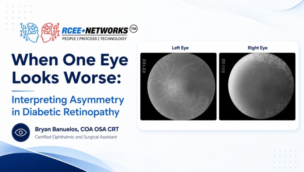
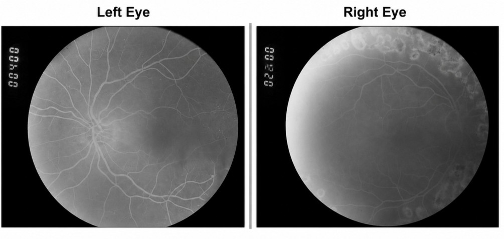
The two retinal images shown here belong to the same patient, yet they tell noticeably different stories. This late-phase angiogram of the right eye shows only a few subtle microaneurysms near the superotemporal macula, along with superior and inferior panretinal photocoagulation scars. There is no active diabetic macular edema or neovascularization visible in this image. However, the presence of prior laser treatment raises important questions about the eye’s history: Was it treated for proliferative diabetic retinopathy, or for severe ischemia associated with advanced nonproliferative disease?
A single image cannot provide every answer. When this eye is compared with the fellow eye, however, the bilateral pattern offers valuable context. Because diabetic retinopathy reflects systemic vascular disease, findings and treatment history in one eye may help clinicians better understand the potential risk and progression of disease in the other.
Patients may present with asymmetric diabetic retinopathy—where one eye appears relatively quiet while the fellow eye demonstrates more advanced disease. For example, one eye may show only subtle microaneurysms and mild nonproliferative diabetic retinopathy (NPDR), while the fellow eye reveals evidence of prior proliferative diabetic retinopathy (PDR), including panretinal photocoagulation (PRP) scars. Although the findings may initially seem discordant, they often represent different stages or treatment histories within the same systemic disease process.
Although diabetic retinal changes often affect both eyes, progression is not always symmetrical. Local factors such as prior treatment, retinal perfusion, vascular anatomy, media clarity, image quality, and imaging timing can influence how disease appears from eye to eye. This means clinicians should be cautious about assuming the less affected eye is at low risk of progression.
Studies evaluating diabetic macular edema (DME) have demonstrated that patients initially presenting with unilateral disease may later develop involvement in the fellow eye during follow-up, particularly within the first several years after diagnosis. These findings reinforce the importance of bilateral surveillance and longitudinal imaging rather than relying on a single snapshot in time.¹
Comparing both eyes may help clinicians distinguish isolated findings from broader patterns of progression, treatment response, or vascular risk. A severely affected eye may prompt closer observation of subtle findings in the fellow eye, while differences between the eyes may provide additional context for clinical decision-making.
Ultimately, retinal images tell a broader story when viewed as a pair. One eye may show where disease has been, while the other may offer insight into where it could be going. Understanding these bilateral patterns reminds clinicians and patients alike that diabetic retinopathy management is not simply about treating isolated findings; it is about recognizing trends, monitoring risk, and protecting vision over time. Platforms such as RetinaWiseAI support side-by-side and longitudinal image review, helping clinicians compare findings across both eyes and make more informed decisions over time.
References:
Alsaloum, P., Russell, M. W., Blaga, V., Kuo, B. L., Wu, A. K., Liu, B. J., Valentim, C. C. S., Xu, C. M., Muste, J. C., Kumar, M., & Singh, R. P. (2023). Time to fellow eye involvement in patients with unilateral diabetic macular oedema. Eye, 37(13), 2761–2767. https://doi.org/10.1038/s41433-023-02410-5