Discover how retinal image analysis is transforming modern eye care through the power of AI-powered clinical decision-support. By analyzing high-resolution retinal images with exceptional speed and precision, these advanced tools help detect eye diseases at their earliest stages, improve diagnostic accuracy, and reduce the risk of vision loss. They also streamline ophthalmology workflows, enabling clinicians to make faster, more informed decisions while enhancing patient care.
Key Takeways:
- AI helps to enable faster and earlier retinal disease detection.
- Improves diagnostic accuracy and consistency.
- Automates retinal image analysis and reporting.
- Enhances clinical efficiency and patient care
Vision is one of our most precious senses; yet, millions of people worldwide lose their sight due to the late detection of various retinal diseases—conditions that could often be prevented if identified in time. Diseases like diabetic retinopathy, glaucoma, and age-related macular degeneration (AMD) typically progress gradually, and often show no symptoms or only very mild ones in the early stages.
The Role of AI in Retinal Image Analysis
Here are the bullet points with clear point names:
- Early Detection is Critical: By the time a patient notices vision changes or other eye-related symptoms, irreversible retinal damage may have already occurred, making early detection essential.
- High-Resolution Retinal Imaging: Modern retinal imaging technology captures detailed images of the retina, enabling clinicians to detect subtle abnormalities that may be missed during routine eye examinations.
- Growing Clinical Workload: The increasing number of retinal images generated in healthcare settings has made manual image review a time-consuming and labor-intensive process.
- AI-Powered Retinal Image Analysis: Advanced retinal image analysis combines artificial intelligence with clinical decision-support to identify disease-related retinal features quickly and accurately.
- Faster Identification of High-Risk Patients: These systems help ophthalmologists prioritize patients who require immediate attention, enabling earlier diagnosis and timely treatment.
- Support for Modern Eye Care: Retinal image analysis has become an essential part of preventive ophthalmology, tele-ophthalmology, and large-scale retinal screening programs.
- Improved Clinical Outcomes: By increasing diagnostic efficiency and enabling timely intervention, retinal image analysis helps preserve vision, improve patient outcomes, and support more effective eye care.
What is Retinal Image Analysis?
- Retinal image analysis is a process that involves analyzing digital images of the retina using advanced software algorithms and AI-powered clinical decision-support to detect, measure, and monitor abnormalities within the eye. Instead of relying solely on human analysis, doctors now use intelligent software capable of automatically analyzing retinal structures and identifying signs of disease.
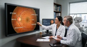
- The retina is a fine layer of tissue at the back of the eye that track the light and transmits visual information to the brain, enabling us to recognize color, shape, and movement. It contains vital structures such as blood vessels, the optic disc, and the macula—which provide valuable clinical information. information regarding the overall health of the eye and the body. Even the slightest change in the retina can be an early warning sign of eye disease, which, if not detected in time, can lead to permanent vision loss.
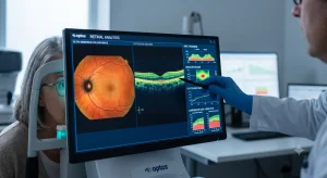
- Retinal image analysis typically utilizes high-quality images obtained through technologies such as color fundus photography,Optical Coherence Tomography (OCT), and OCT Angiography (OCTA). The software analyzes these images to detect issues such as blood vessel abnormalities, retinal lesions, changes in the optic nerve, fluid accumulation, and macular degeneration.
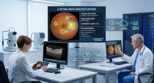
- Modern AI-powered clinical decision-support systems can analyze these images and identify suspicious areas within seconds. They can determine the severity of a disease and generate structured reports, thereby assisting ophthalmologists in making accurate clinical decisions. Rather than replacing doctors, these tools boost confidence in disease diagnosis and enhance treatment process.
Why Retinal Image Analysis Is Important?
The demand for retinal image analysis has risen significantly as eye diseases become increasingly common worldwide. Global health estimates indicate that millions of people living with diabetes are at risk of diabetic retinopathy, while an aging population is increasingly susceptible to glaucoma and age-related macular degeneration.
Provide retinal image analysis offers several key benefits:
1. Detects diseases at an early stage Improves diagnostic consistency
2. Reduces clinician workload Enables large-scale screening programs
3. Supports remote eye care via tele-ophthalmology
4. Enhances patient monitoring over time
5. Improves treatment planning through quantitative measurements
Also Read:AI Retinal Image Analysis Tools
Detecting Disease Through Retinal Image
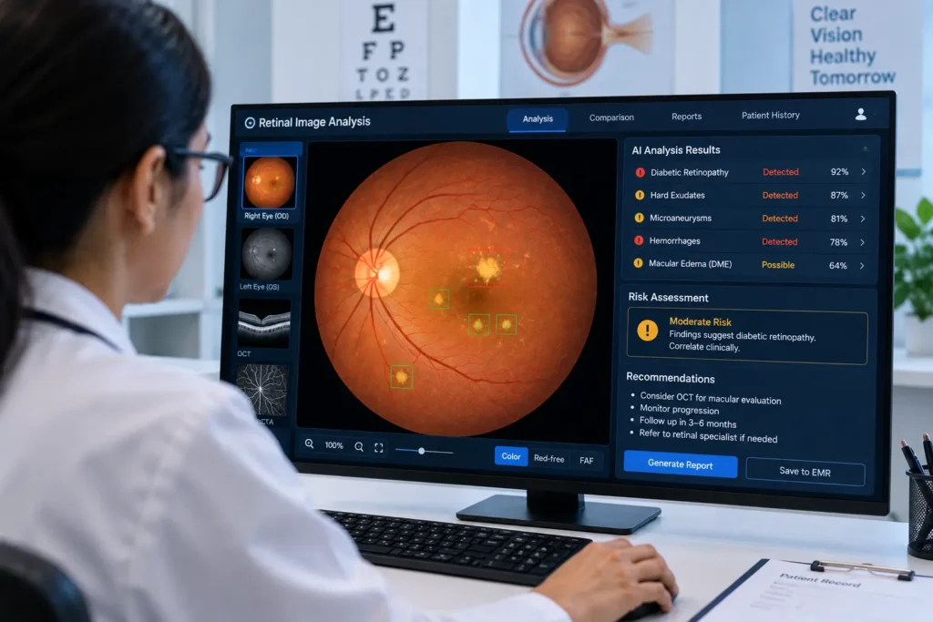
Analysis Retinal image analysis has become a key tool in diagnosing and keeping track of a wide range of eye conditions, One of the most useful thing of this technology is detecting diabetic retinopathy. AI-powered clinical decision-support systems can identify microaneurysms, hemorrhages, cotton-wool spots, and hard exudates, making it possible to step in early before serious vision loss develops.
Age-related macular degeneration affects the central part of the retina, resulting in significant vision loss. Image analysis helps detect drusen deposits, pigmentary abnormalities, and fluid buildup that are associated with the disease getting worse over time.
Glaucoma Slowly damages the optic nerve, often without any particular symptoms in the primary stages. Retinal image analysis can measure the optic cup-to-disc ratio and the thickness of the retinal nerve fiber layer, giving clinicians valuable information for catching glaucoma before it progresses too far.
Process of Retinal Imaging
- Beyond these conditions, the technology is also useful for identifying hypertensive retinopathy, retinal vein occlusion, macular edema, optic disc abnormalities, and various inherited retinal diseases.
- By monitoring patients over time, clinicians can get a clearer picture of how a disease is progressing and how well a given treatment is working. AI-Powered Clinical Decision-Support and Deep Learning in Retina Image Analysis. Artificial intelligence has transformed retinal image analysis from a mere supportive tool into an intelligent clinical assistant.
- Convolutional Neural Networks (CNNs)—one of the most important models in deep learning technology—are trained using millions of retinal images. Through this extensive training, the model can accurately identify the subtle and complex characteristics of various eye diseases and deliver highly precise results.
- These AI-powered clinical decision-support systems can automatically classify retinal diseases, segment anatomical structures, detect microscopic lesions, and prioritize urgent cases for expert review. Techniques such as heatmaps and explainable AI enhance transparency by revealing the specific areas of the image that influenced the model’s predictions.
- Rather than replacing ophthalmologists, AI augments their expertise by reducing repetitive tasks, minimizing inter-observer variability, and facilitating faster clinical decision-making. This is particularly valuable in regions facing a shortage of ophthalmologists, where clinical decision-support-assisted screening can expand access to high-quality eye care.
Retinal image analysis is more than just a technological advancement—it is a crucial step toward safeguarding the gift of sight. By facilitating early disease detection and timely treatment, it helps preserve vision and improve the people’s quality of life. As AI-powered clinical decision-support technologies and medical imaging continue to evolve, they are shaping a future where preventable blindness is becoming increasingly rare.
Frequently Frequently Asked Questions (FAQs)
1.What is retinal image analysis?
Retinal image analysis uses software and AI-powered clinical decision-support to evaluate retinal images, helping detect and monitor eye diseases more efficiently.
2.Which diseases can retinal image analysis detect?
It can identify diabetic retinopathy, glaucoma, age-related macular degeneration, hypertensive retinopathy, macular edema, retinal vein occlusion, and other retinal abnormalities.
3.Does AI replace ophthalmologists?
No. AI-powered clinical decision-support supports ophthalmologists by providing faster image analysis and decision support, while final diagnosis and treatment decisions remain with qualified clinicians.
4.What types of retinal images are commonly analyzed?
The most common modalities include color fundus photography, Optical Coherence Tomography (OCT), OCT Angiography (OCTA), and fluorescein angiography.
5.Why is retinal image analysis important?
It enables earlier disease detection, improves diagnostic consistency, enhances clinical efficiency, and supports timely treatment, ultimately helping preserve patients’ vision.