RCEE+Networks LLC
  • Home
  • About
  • Services
    • IMACAM™ Tech Support
  • Products
    • RetinaWiseAI™
  • Contact
+1-844-2-THEOWL
info@rceenetworks.com
logotype
  • Home
  • About
  • Services
    • IMACAM™ Tech Support
  • Products
    • RetinaWiseAI™
  • Contact
logotype
  • Home
  • About
  • Services
    • IMACAM™ Tech Support
  • Products
    • RetinaWiseAI™
  • Contact
Health
Home Archive by Category "Health"

Category: Health

EducationHealthLearning
Umesh Kumar KhiriNovember 14, 2025 0 Comments
Share article:TwitterFacebookLinkedin
27 Views
11 Likes

RETINAL IMAGE ANALYSIS USING AI TECHNOLOGY AT RCEENETWORKS

Retinal image analysis using AI technology

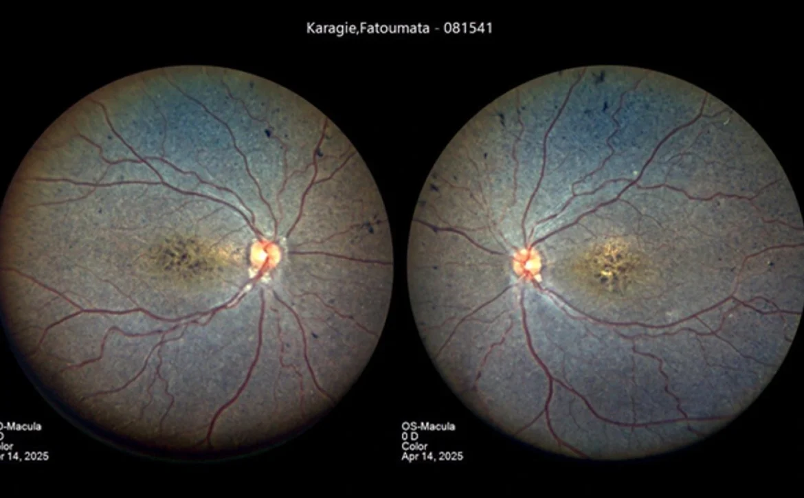
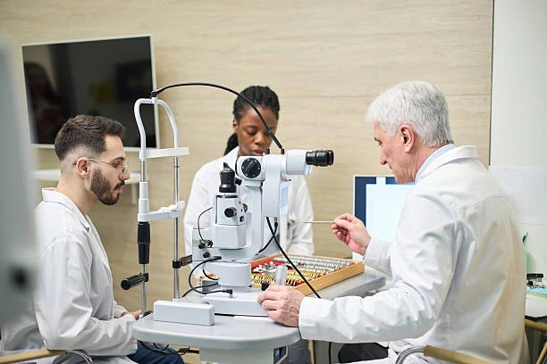
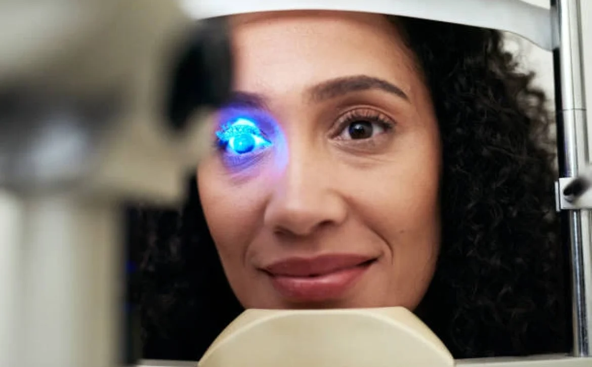
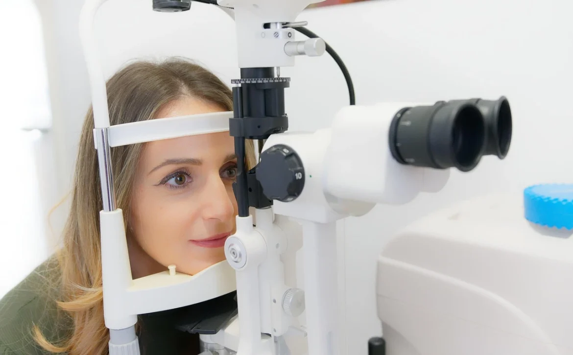
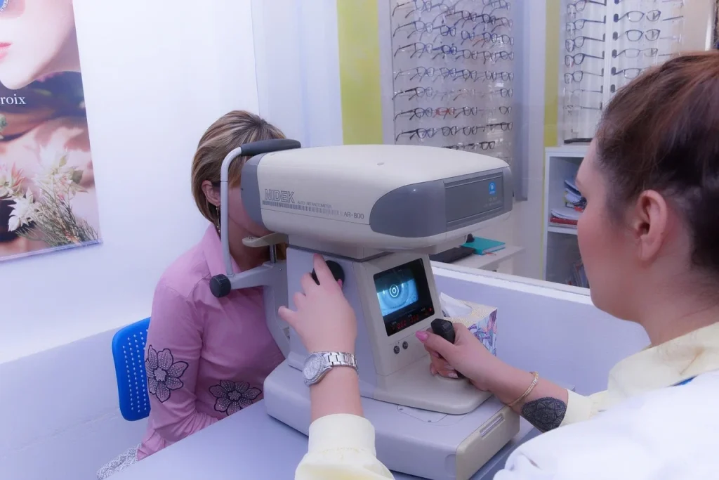
Image analysis for retinal images has been an active subject of research over the last couple of decades. With the popularity of mydriatic and non-mydriatic digital imaging cameras, color fundus photographs have become essential part of standard retinal exam. Advances in image analysis, pattern recognition, and machine learning have opened up a great opportunity to enhance the clinical care available to patients suffering for retinal diseases such as diabetic retinopathy, age related macular degeneration, hypertensive retinopathy, and glaucoma.

Rceenetworks logo

RCEENetworks’ Optometry & Ophthalmology software boosts accuracy, efficiency, and patient care for eye care professionals.

Eye care professional showing a patient a report beside an Optos imaging machine.

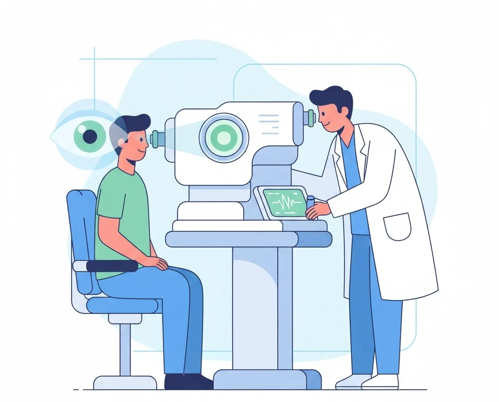
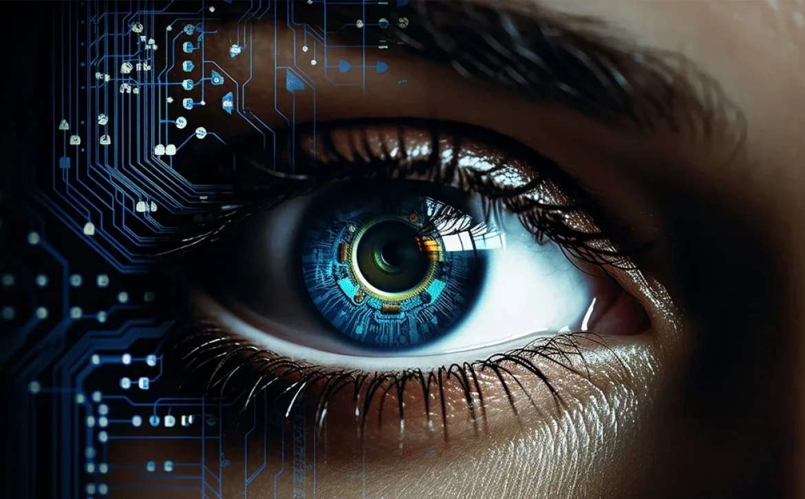
RCEENETWORKSTM is developing exciting new technologies to detect various lesions related to retinal diseases such as diabetic retinopathy (DR), age related macular degeneration (ARMD), Glaucoma, Edema – to name a few. RCEENETWORKSTM engineers have deep understanding of image processing theory and have leveraged this experience to create intelligent AI models. In addition, they combine the AI image analysis output with patient’s medical data to increase diagnostic accuracy to the highest level. Watch this space for the release of our next generation imaging analysis system – RetinaWiseAI .
Retinal image analysis using AI technology

READ MORE
HealthLearning
Umesh Kumar KhiriNovember 13, 2025 0 Comments
Share article:TwitterFacebookLinkedin
27 Views
14 Likes

Revolutionizing Vision Care: AI-Powered Diagnosis of Rod-Cone Dystrophy

Diagnosis: Cone-Rod Dystrophy (CRD)

Observed Features:

  • Macular pigmentary changes (mottling and granularity) in both eyes.
  • Slightly reduced fundus view clarity.

Reasoning:

Quality Assessment: The image quality is slightly grainy and has a somewhat washed-out appearance, possibly indicating mild media opacity like a subtle cataract, which marginally reduces fundus clarity.

Systematic Inspection:

Optic Disc Examination:

The optic discs appear normal in terms of cup-to-disc ratio and neuroretinal rim. No signs of optic disc hemorrhages or notching are visible.

Macular Examination:

In both eyes, there is a noticeable abnormality in the macula. We observe pigmentary changes described as mottling and granularity within the macular region. There is no clear evidence of drusen, hemorrhages,or exudates typically associated with AMD or DR. Macular edema is not suggested by the absence of macular thickening or cystoid spaces.

Retinal Vessel Examination:

The retinal vessels appear generally normal. There are no signs of microaneurysms, hemorrhages, cotton wool spots, venous beading, IRMA, or neovascularization, which are key features of DiabeticRetinopathy. Vessel caliber seems within normal limits.

Differential Diagnosis:

Diabetic Retinopathy (DR):

Ruled out due to the absence of microaneurysms,hemorrhages, exudates, and neovascularization. Age-Related Macular Degeneration (AMD): Less likely as typical drusen are not clearly observed. While pigmentary changes are present, the pattern is more of a diffuse mottling and granularity rather than the typical drusen or geographic atrophy of AMD. Wet AMD features are absent.

Glaucoma:

Ruled out as the optic discs appear normal, with no signs of increased CDR, rim thinning, or disc hemorrhages.

Macular Edema:

Not indicated due to the lack of macular thickening, cystoid spaces, or associated exudates.

Cataract:

Mild cataract may be present, indicated by the slight reduction in image clarity, but it’s not the primary finding.

Cone-Rod Dystrophy (CRD):

The observed macular pigmentary changes, specifically the mottling and granularity, are consistent with macular atrophy, a key feature of Cone-Rod Dystrophy. This, in the absence of strong indicators for other conditions, makes CRD the most probable diagnosis based on these images.

Conclusion:

Based on the macular pigmentary changes (mottling and granularity) and the exclusion of other more common retinal conditions based on feature absence, Cone-Rod Dystrophy is the most likely diagnosis. The mild reduction in fundus clarity could be due to a subtle cataract, but this is considered a secondary finding compared to the macular pathology.
Courtesy – Dennis West.

READ MORE
Health
Umesh Kumar KhiriNovember 6, 2025 0 Comments
Share article:TwitterFacebookLinkedin
29 Views
8 Likes

RCEENETWORKS LLC – AI-powered retinal image analysis

Key Features:

  • Ensures GDPR compliance with secure and pseudonymized patient data processing.Utilizes advanced deep learning algorithms for precise retinal image assessment.
  • Delivers analysis results in approximately one minute.
  • Aligns with international standards, including ICDR and AREDS.
  • Compatible with all major fundus cameras and adaptable to various clinical workflows.

Table of Contents

  • Key Features:
  • RetinaWiseAI
  • Revolutionizing retinal eye care with AI-powered image analysis
  • Harnessing AI for better outcomes
  • Efficient workflow with RetinaWiseAI
  • Seamless integration
    • Speeding up your workflow
    • Early Detection of Retinal Diseases
    • Instrument Integration

RetinaWiseAI

RetinaWiseAI is an AI-powered software solution designed to analyze color fundus images for the early detection of vision-threatening conditions such as diabetic retinopathy (DR), age-related macular degeneration (AMD), and glaucoma. This solution leverages advanced deep learning and computer vision technologies to provide accurate and reliable analysis results.

Revolutionizing retinal eye care with AI-powered image analysis

RetinaWiseAI transforms retinal disease detection by harnessing AI-driven image analysis to support healthcare professionals in identifying vision-threatening conditions. It delivers accurate, reliable results that enhance clinical decision-making.
By integrating detail-rich confocal fundus imaging and cutting-edge AI-powered retinal image analysis, the RCEENETWORKS screening solution streamlines workflows, enhances diagnostic accuracy, and improves the patient care journey.

Harnessing AI for better outcomes

Retinal diseases account for up to 80% of preventable blindness cases, with global vision loss costs approaching $3 trillion annually. Early detection is critical. RCEENETWORKS’s AI-driven analysis transforms retinal image interpretation by delivering intuitive reports that help healthcare professionals make informed decisions quickly and accurately, improving patient outcomes and engagement.

Efficient workflow with RetinaWiseAI

Optometrist performing eye examination using slit lamp on a patient.

Quick image capture:
Digital color fundus cameras acquire images in seconds.
Automated analysis:
AI processes images, generating scores and heat maps requiring only few seconds of computing time
GDPR compliance:
Patient data is pseudonymized for secure handling.
Accessible reports and EMR Integration:
Results integrate directly into existing EMR software.

Seamless integration

RetinaWiseAI is compatible with all major fundus camera vendors, ensuring versatility in diverse clinical and optical settings. While its adaptability extends to various imaging devices. RetinawiseAI redefine the possibilities of remote retinal screening, offering consistent performance and better patient care.
With RetinaWiseAI, eye care specialists gain an advanced tool for safeguarding vision, streamlining workflows, and enhancing the patient care journey—while the final diagnosis remains in the hands of the doctor.

Speeding up your workflow

With artificial intelligence, healthcare organizations can conduct large-scale eye screenings efficiently and accurately. RCEENETWORKS deliver results that match—or even surpass—traditional vision screening methods in reliability. By instantly analyzing every patient’s retinal image using clinically validated software trained to detect disease patterns, RetinaWiseAI empowers fast, precise, and scalable screening for entire populations.

Early Detection of Retinal Diseases

Using artificial intelligence, opticians can now detect early signs of age-related macular degeneration, diabetic retinopathy and glaucoma, enabling faster diagnosis. RetinaWiseAI, combined with fundus imaging, delivers reliable and consistent performance regardless of workload or time of day. With AI-powered precision, disease detection takes only a few minutes, providing validated, actionable results instantly.

Instrument Integration

RetinaWiseAI seamlessly integrates into clinic-specific workflows, reducing workloads for certified eye care specialists and enhancing clinical capacity. By enabling faster and objective prioritization of patients requiring immediate follow-up RCEENETWORKS LLC transforms eye care. Ophthalmologists can now identify severe symptoms more efficiently, helping more patients receive timely, life-changing care.

READ MORE
HealthEducation
Optometrist checking patient eyesight
Umesh Kumar KhiriNovember 4, 2025 0 Comments
Share article:TwitterFacebookLinkedin
32 Views
10 Likes

Making Data Work for You: Advancing Your Ophthalmic Workflow with RetinaWiseAI

In today’s data-driven world, healthcare professionals are increasingly turning to smart technology to streamline their practices, enhance accuracy, and improve patient outcomes. Ophthalmology is no exception. With the rise of artificial intelligence (AI) and machine learning, innovative tools like RetinaWiseAI are transforming the way eye care professionals diagnose, treat, and manage retinal diseases.

Table of Contents

  • The Growing Challenge in Ophthalmology
  • Ophthalmic Workflows
  • Key Features:
  • How RetinaWiseAI Advances Your Workflow
  • Real-World Impact
  • Conclusion: The Future Is Clearer with RetinaWiseAI

The Growing Challenge in Ophthalmology

Age-related macular degeneration (AMD), diabetic retinopathy, and other retinal diseases are becoming more prevalent, particularly in aging populations. Early detection is critical, yet traditional diagnostic workflows often rely on time-consuming manual analysis and subjective interpretations.

Ophthalmologists are inundated with patient data — fundus images, OCT scans, medical histories — all of which require fast, precise interpretation. This pressure has increased the demand for smart, scalable solutions that can handle large volumes of complex data while reducing clinician workload.

Ophthalmic Workflows

RetinaWise AI is an AI-powered platform designed specifically for ophthalmic professionals. By leveraging deep learning algorithms trained on thousands of retinal images, RetinaWiseAI delivers rapid and accurate insights directly to clinicians, helping them detect early signs of disease with unprecedented speed and confidence.

Key Features:

Automated Image Analysis

Instantly analyzes OCT and fundus images for abnormalities such as drusen, fluid buildup, or retinal thinning.
Risk Stratification
Flags patients based on severity and urgency, helping clinicians prioritize care.
Workflow Integration
Seamlessly fits into existing EMR and imaging systems, reducing the need for additional manual steps.
Decision Support
Offers second-opinion AI insights to support clinical judgment, not replace it.

How RetinaWiseAI Advances Your Workflow

Speeds Up Diagnosis
AI-based triage enables ophthalmologists to focus on critical cases first, reducing diagnostic delays.
Reduces Errors
Standardized image interpretation helps minimize variability between clinicians, leading to more consistent care.
Optimizes Resources
Clinics can handle a higher volume of patients without overburdening their staff, improving overall efficiency.
Supports Teleophthalmology
RetinaWiseAI makes remote diagnostics more reliable, helping rural and underserved populations access expert care.

Real-World Impact

Ophthalmologists conducting an eye examination using slit-lamp equipment.

Clinics using RetinaWise AI have reported:

  • 30–50% reduction in diagnostic time
  • Improved early detection rates for AMD and diabetic retinopathy
  • Higher patient satisfaction through faster consultations
  • Much better Ophthalmic Workflows

Conclusion: The Future Is Clearer with RetinaWiseAI

RetinaWise AI is more than just a diagnostic tool — it’s a powerful ally in the fight against vision loss. By putting data to work intelligently, it empowers eye care providers to make faster, more accurate decisions and ultimately offer better outcomes for their patients.

If you’re looking to modernize your ophthalmic workflow and stay ahead of the curve, RetinaWise AI designed by RCEENetworks is the solution designed with your needs in mind.

READ MORE
HealthEducation
Umesh Kumar KhiriOctober 31, 2025 0 Comments
Share article:TwitterFacebookLinkedin
34 Views
10 Likes

AI Trends and Developments in Optometry Practice Management

AI use in optometry is set to usher in a transformation for the future of eyecare practice management.

Various AI-powered technologies are revolutionizing practice management areas such as appointment scheduling, inventory management, and clinical decision-making.

AI technologies (RetinaWise AI) developed by RCEENetworks, such as deep learning algorithms and convolutional neural networks (CNNs), are being utilized to diagnose and manage various eye conditions, including glaucoma, diabetic retinopathy, and age-related macular degeneration (AMD).

These AI systems can analyze vast amounts of data from retinal images, providing optometrists with precise and timely insights that improve patient outcomes.

Understanding how artificial intelligence is poised to reshape optometry practices can lead to streamlined operations, increased efficiency and accuracy, and enhanced patient care.

Table of Contents

  • AI Applications in Optometry Practice Management
  • Chatbots and Large Language Models (LLMs)
  • AI Tools for Eye Examinations
    • AI-powered vision correction:
    • AI-driven eye diagnostics:
    • AI-assisted ocular imaging:
    • AI for early glaucoma detection:
  • AI in Teleoptometry
    • Algorithms for Optometry Patient Feedback
  • Move Your Practice Into the Future with Cloud-Based Technology

AI Applications in Optometry Practice Management

AI applications are changing how optometrists implement practice management, improving patient care and boosting efficiency. For instance, AI-powered technology improves scheduling, customer service, and practice performance tracking.

Chatbots and Large Language Models (LLMs)

The integration of Artificial Intelligence (AI) and Large Language Models (LLMs) in optometry is revolutionizing the field, enhancing both diagnostic accuracy and patient care.

These AI-driven virtual assistants can improve patient interactions within optometry practices. Capable of handling routine inquiries and frequently asked questions, chatbots provide real-time responses and support to patients, even outside regular office hours.

LLMs, such as GPT-4, are also making significant contributions to optometry. These models can assist in medical transcription, enhance electronic health records, and provide clinical decision support.

Their conversational interfaces simulate human-like interactions, creating a personalized experience for patients seeking information about services, insurance, or general eye care advice. This instant and continuous support increases patient engagement and improves the practice’s overall responsiveness.

Recent studies have shown that LLMs can match or even outperform ophthalmologists in diagnosing and recommending treatments for conditions like glaucoma and retinal diseases.

RCEENetworksTweet

Furthermore, LLMs are being incorporated into medical education, helping to train the next generation of eye care professionals by familiarizing them with emerging technologies.

As AI technologies get even better, chatbots are evolving into even ever-more realistic service assistants to further improve patient outcomes and streamline optometrists’ workflows.

AI Tools for Eye Examinations

an ophthalmologist performing an eye exam using diagnostic equipment on a patient.

Optometrists are using emerging AI tools to improve eye examinations and provide more accurate patient treatments. Tools that are available now include AI-powered vision correction tools, diagnostics, imaging, and glaucoma detection.

One notable advancement is the development of AI tools like RetinWise AI, which exemplifies how AI can assist in early detection and management of eye diseases, potentially preventing vision loss in patients.

Additionally, AI-driven platforms like RetinaWise AI are being used in clinical settings to screen and grade eye conditions, aiding optometrists in making informed decisions about treatment plans.

AI-powered vision correction:

AI technologies improve vision correction by precisely analyzing patient data, leading to personalized prescriptions for optimal visual acuity. This ensures a more tailored and precise eyewear experience.

AI-driven eye diagnostics:

Through advanced algorithms, AI aids optometrists in diagnosing eye conditions with greater accuracy and efficiency. The analysis of patient data enables early detection and more effective treatment plans. For example, AI algorithms in optometry have led to better prediction, diagnosis, screening, and treatment for myopia.

AI-assisted ocular imaging:

AI enhances ocular imaging by providing in-depth insights into retinal health and other eye structures. This assists in identifying potential ocular issues, leading to proactive eye care measures and advances in ophthalmological treatment.

AI for early glaucoma detection:

AI’s capabilities enable early prediction of glaucoma incidence and progression, helping eyecare providers diagnose and treat the condition early on. In a 2022 study, the AI model used to predict glaucoma showed a high accuracy of 90% in the validation set and remained consistent, with 89% and 88% accuracy in two external test sets.

By analyzing relevant data and retinal photographs, AI supports timely intervention, promoting better management of glaucoma and preserving vision.

AI in Teleoptometry

AI and digital technology integration in ophthalmology with data analytics, robotics, and cloud health icons.

AI-driven tools are transforming telehealth in optometry, bringing better patient services, efficiency, and accuracy to eye care.

Optometrists can provide remote consultations and eyecare screenings using virtual platforms powered by artificial intelligence. This enables patients to connect with optometrists from the comfort of their homes or remote locations.

As part of a remote eye care appointment, patients can capture images of their retina using a smartphone or specialized retinal camera. When providing telemedicine for retinal care, providers can use AI-powered tools to analyze images for signs of eye disease, such as diabetic retinopathy, macular degeneration, or glaucoma.

For instance, the Navilas® Laser System, a navigated laser photocoagulator for ophthalmology, allows retina specialists to preplan treatments at specific locations and times. This tool combines laser photocoagulation with fluorescein angiographic imaging, achieving an impressive 92% accuracy of hitting microaneurysms in diabetic macular edema (DME) compared to the standard manual technique laser treatment, with 72% accuracy.

Algorithms for Optometry Patient Feedback

AI tools are transforming how optometry practices gather and use patient feedback. Through natural language processing, sentiment analysis, and data mining, AI can process vast amounts of patient feedback from various sources, such as surveys and online reviews. This allows optometrists to gain valuable insights into patient experiences, preferences, and concerns.
With AI’s ability to identify patterns and trends, optometry practices can pinpoint areas for improvement and implement targeted enhancements to enhance patient satisfaction.

Move Your Practice Into the Future with Cloud-Based Technology

Overall, AI is transforming optometry, offering new tools and capabilities that enhance the accuracy, efficiency, and quality of eye care.

Not unlike AI in optometry, cloud-based RevolutionEHR is revolutionizing eye care. RetinaWise AI, by RCEENetworks offers the all-in-one functionality, streamlined workflows, and seamless partner integrations today’s optometrists need.

Embracing cloud-based technologies empowers optometrists to provide efficient and patient-centered treatments, ultimately shaping the future of eye care.

READ MORE
Health
Umesh Kumar KhiriOctober 29, 2025 0 Comments
Share article:TwitterFacebookLinkedin
35 Views
10 Likes

Unlocking Efficiency in Ophthalmology ASCs with RetinaWise AI

In ophthalmology, every surgical day runs on tight schedules and tighter margins. A single missed clearance, outdated allergy list, or unconfirmed plan can ripple across an entire surgical block. This derails case flow, wastes staff time, and frustrates patients.

Yet for many ASCs, surgical coordination still relies on a fragile mix of paper packets, spreadsheets, email threads, and verbal updates. Staff manually re-enter data between systems. Tasks are tracked in silos. And coordination depends more on memory and hallway conversations than on system reliability.

This isn’t just inefficient. It’s unsustainable.

Table of Contents

  • The Cost of Surgical Inefficiency
  • Where Current Systems Fall Short
  • What True Integration Looks Like
  • Why Integration Matters
  • Introducing RetinaWise AI
  • The Gains from AI-Driven Integration
  • Why RetinaWise AI is Different
  • Build a Scalable Future for Your ASC

The Cost of Surgical Inefficiency

Surgical days should run on precision, but for many ophthalmology practices, they run on patchwork. Coordinators print out paper forms. Nurses check with the front desk about clearances. Surgical techs search email threads or spreadsheets for missing information.

These manual workarounds aren’t just inefficient—they create risk. The disconnect between systems forces staff to double-check what should already be clear, slowing down patient flow and increasing pressure across the board.

In ophthalmology, where surgical volume is high and timelines are tight, the margin for error is slim. Each denied surgical claim can cost $300–$600 to rework. With a 5–10% denial rate due to documentation issues, these inefficiencies have real financial impact.

Where Current Systems Fall Short

Most ASCs use a combination of EHRs, spreadsheets, paper packets, and phone calls to coordinate care. These systems rarely talk to each other. When data needs to be shared, it’s re-entered manually or confirmed verbally—introducing errors.

  • This fragmentation means:
  • No single source of truth for the surgical schedule
  • Staff forced to track details by memory or silos
  • Surgeons walk into cases with incomplete or outdated plans
  • Nurses delay prep while waiting for verbal confirmation
  • Schedulers rearrange blocks manually like a game of Tetris

As surgical volume grows, cracks in the system widen. Without integration, ASCs hit a ceiling—they cannot scale without overwhelming their teams.

What True Integration Looks Like

Integration means systems that share data instantly and accurately across departments. In a fully integrated ASC:

  • Data entered once populates everywhere it’s needed
  • Clearances sync automatically from the EHR to the surgical schedule
  • Consent forms, pre-op readiness, and post-op notes update in real time
  • Clinics can submit ASC scheduling requests directly

Instead of people acting as bridges between tools, the system itself ensures clarity, visibility, and accountability.

Why Integration Matters

When workflows are fragmented, the costs are real:

  • Staff burnout rises as nurses and admins chase paperwork instead of patients
  • Patient experience suffers from delays, repeated questions, and disorganization
  • Revenue is lost every time a case is delayed, canceled, or underutilized

You can’t grow surgical volume on a workflow that breaks under pressure. Integration is the infrastructure your ASC needs to deliver precision at scale.

Introducing RetinaWise AI

A woman is seated at an eye exam machine, adjusting her position for a vision test.

RetinaWise AI is designed to solve these very challenges for ophthalmology ASCs. Unlike generic tools, it’s built for the unique demands of cataract, retina, glaucoma, and corneal surgery workflows.

  • With RetinaWise AI, practices can:
  • Eliminate redundant data entry across systems
  • Ensure clearances, consents, and surgical plans sync automatically
  • Gain real-time visibility into every case’s readiness
  • Track pre-op, intra-op, and post-op workflows from one interface
  • Reduce admin overhead while increasing throughput

The Gains from AI-Driven Integration

Adopting RetinaWise AI produces measurable improvements:

  • Recovered Lost Case Volume – Prevent cancellations and delays caused by missing documentation, recapturing up to $150,000 annually.
  • Increased Throughput Without Adding Staff – Add 1–2 more cases per OR day, driving $400,000–$800,000 in additional revenue.
  • Reduced Admin and Overtime Costs – Save 8–10 hours per week of manual tracking and coordination, freeing staff to focus on patients.
  • Improved Surgeon Efficiency – Minimize idle time, protecting $75,000–$100,000 annually in lost clinical productivity.

Why RetinaWise AI is Different

  • Purpose-built for ophthalmology – Designed around real-world surgical workflows, not generic templates
  • Seamless integration – Connects with EHR and admin systems for end-to-end workflow visibility
  • Rapid adoption – Built to match how ophthalmology staff already work, reducing training time
  • Scalable by design – Supports higher surgical volumes without burning out staff
  • This isn’t just an efficiency upgrade—it’s the foundation for scalable, reliable surgical operations.

Build a Scalable Future for Your ASC

For ophthalmology practices, ASC integration isn’t optional. It’s the difference between running at 80% capacity with constant friction—or running at full precision, with room to grow.

RetinaWise AI provides the infrastructure for surgical reliability, efficiency, and growth.
Learn more about how RetinaWise AI can transform your surgical workflows: https://retinawiseai.com/ or Request a Free Demo

READ MORE
Health
Umesh Kumar KhiriApril 4, 2021 2 Comments
Share article:TwitterFacebookLinkedin
503 Views
49 Likes

Connecting The Business Technology Community

In order to write the perfect blog post, you need to break your content up into paragraphs. While most blog posts use paragraphs, few use them well. Take the time to put links in your blog post…

READ MORE
Health
Umesh Kumar KhiriSeptember 23, 2020 2 Comments
Share article:TwitterFacebookLinkedin
444 Views
50 Likes

Transforming the way the world learns

In order to write the perfect blog post, you need to break your content up into paragraphs. While most blog posts use paragraphs, few use them well. Take the time to put links in your blog post…

READ MORE
Health
Umesh Kumar KhiriSeptember 23, 2020 2 Comments
Share article:TwitterFacebookLinkedin
346 Views
36 Likes

Combating Ransomware for a New Era of Work

In order to write the perfect blog post, you need to break your content up into paragraphs. While most blog posts use paragraphs, few use them well. Take the time to put links in your blog post…

READ MORE

Recent Posts

  • RETINAL IMAGE ANALYSIS USING AI TECHNOLOGY AT RCEENETWORKS
  • Revolutionizing Vision Care: AI-Powered Diagnosis of Rod-Cone Dystrophy
  • AI makes retinal imaging 100 times faster, compared to manual method
  • Artificial intelligence: the Unstoppable Revolution in Optometry and Ophthalmology
  • Progress in AI for Retinal Image Analysis

Recent Comments

  1. Fred Hyman on Yellow Cap
  2. Fred Hyman on T-Shirt Full Hand
  3. Fred Hyman on Yellow Cap
  4. Randal Gray on Black T-Shirt
  5. John Harris on Black T-Shirt

Archives

  • November 2025
  • October 2025
  • April 2021
  • March 2021
  • September 2020
  • April 2020

Categories

  • Business
  • Education
  • Finance
  • Health
  • Learning
  • Lifestyle
  • Students
  • Uncategorized
Recent Posts
  • RETINAL IMAGE ANALYSIS USING AI TECHNOLOGY AT RCEENETWORKS
    RETINAL IMAGE ANALYSIS USING AI TECHNOLOGY AT RCEENETWORKS
    November 14, 2025
  • Revolutionizing Vision Care: AI-Powered Diagnosis of Rod-Cone Dystrophy
    Revolutionizing Vision Care: AI-Powered Diagnosis of Rod-Cone Dystrophy
    November 13, 2025
  • AI makes retinal imaging 100 times faster, compared to manual method
    AI makes retinal imaging 100 times faster, compared to manual method
    November 11, 2025
Categories
  • Business2
  • Education6
  • Finance5
  • Health9
  • Learning4
  • Lifestyle3
  • Students3
  • Uncategorized4
Tags
balance coach coaching courses education eye care lifeguide motivation Ophthalmic Ophthalmology optometrist strategy
logotype

+1-844-2-THEOWL (+1 844 284-3695)‬

info@rceenetworks.com

3010 LBJ Freeway, Suite 1200, Dallas, Texas 75234

Menu

Home Services

ProductBlog

Contact

Other Links

RetinaWise AIPrivacy PolicyContent PolicyTerms & Conditions

Social Links

TwitterFacebookInstagramLinkedin

Copyright © 2025 RCEENetworks LLC. All Rights Reserved