When reviewing a fluorescein angiogram (FA), it’s easy to focus on the late frames where leakage becomes most apparent. However, in retinal vein occlusions (RVOs), some of the most valuable information is found within the first few seconds of the study.
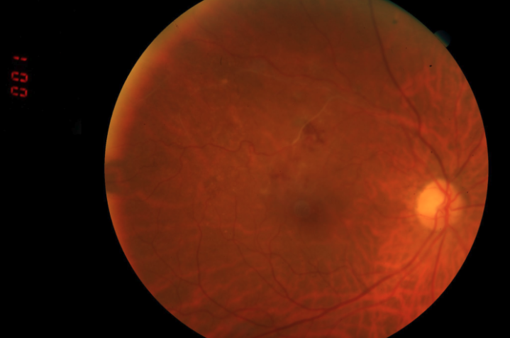
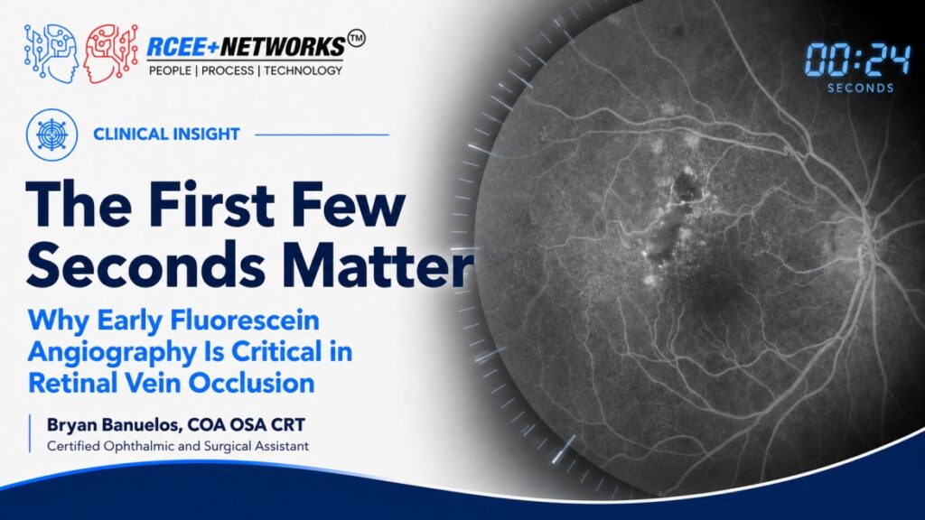
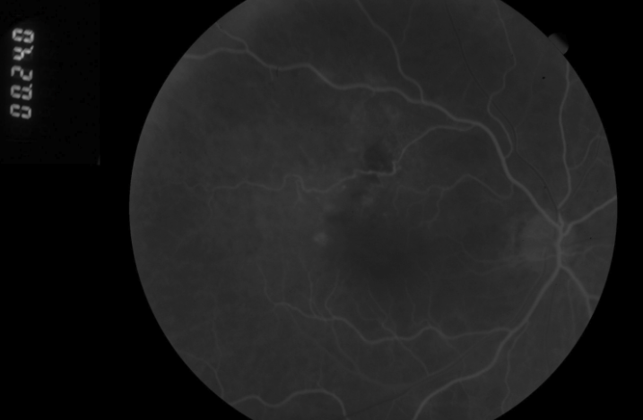
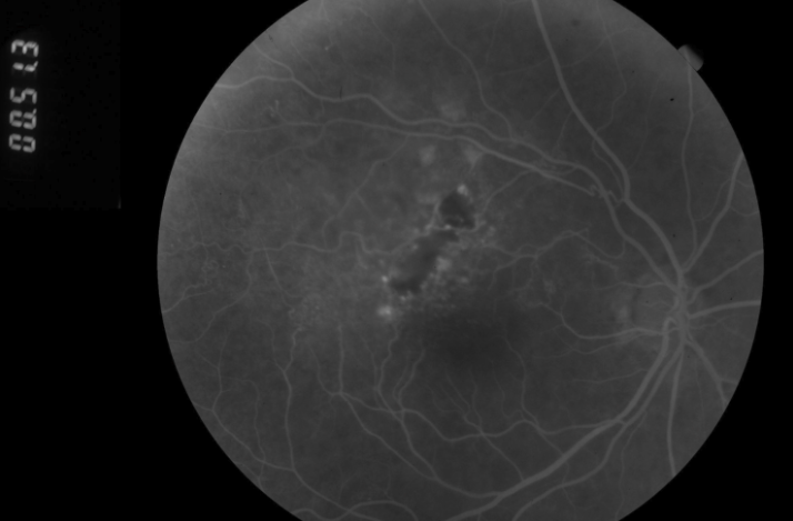
We recently evaluated a patient with a superotemporal branch retinal vein occlusion (BRVO) of the right eye. Early-phase FA demonstrated a markedly prolonged arteriovenous (AV) transit time, along with a ghost venule supplying an area of macular capillary nonperfusion. The angiogram provided a striking example of how delayed venous drainage correlates with irreversible retinal ischemia.
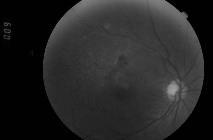
AV transit time is an important marker when distinguishing ischemic from non-ischemic RVOs. While non-ischemic occlusions often maintain relatively preserved retinal perfusion, ischemic RVOs typically demonstrate severely delayed venous filling accompanied by extensive capillary nonperfusion. These ischemic areas become hypoxic, stimulating the release of vascular endothelial growth factor (VEGF) and increasing the risk of neovascularization.
Recognizing these findings early is essential because they help us risk stratify patients for complications such as retinal neovascularization, vitreous hemorrhage, and, in severe cases, neovascular glaucoma. Identifying significant ischemia also guides how closely a patient should be monitored during follow-up.
Unfortunately, there is currently no treatment proven to restore retinal capillary perfusion once macular ischemia has developed. Management instead focuses on treating associated macular edema when present, monitoring carefully for neovascularization, and intervening promptly if new vessels develop.
Anti-VEGF therapy remains the standard of care for RVO-associated macular edema and is highly effective in treating neovascular complications. However, laser photocoagulation continues to play an important role in eyes with significant ischemia and high-risk neovascularization. Working alongside a second-generation retina specialist has given me the opportunity to appreciate a practice philosophy that often favors laser treatment in these cases—a strategy supported by decades of clinical experience. As with many retinal diseases, management is ultimately individualized based on each patient’s clinical and angiographic findings.
This case serves as a great reminder that fluorescein angiography isn’t just about identifying leakage. Sometimes the most clinically significant information is captured before the leakage even begins. Careful attention to early-phase imaging and AV transit time can provide invaluable prognostic information and help guide long-term management in patients with retinal vein occlusions.
Further Reading
American Academy of Ophthalmology. Retinal Vein Occlusions Preferred Practice Pattern®.
American Academy of Ophthalmology. “Branch Retinal Vein Occlusion.” EyeWiki.
American Society of Retina Specialists. “Branch Retinal Vein Occlusion.”