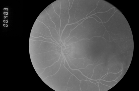
As retinal imaging becomes more advanced, clinicians are identifying diabetic retinopathy earlier than ever before. The challenge is no longer simply recognizing disease. It is determining when early findings warrant treatment versus careful observation. While early detection remains critical for preserving long-term vision, identifying disease does not always mean treatment must begin immediately.
Diabetic retinopathy develops when chronic hyperglycemia damages retinal blood vessel function. In the early stages, known as nonproliferative diabetic retinopathy (NPDR), findings may include microaneurysms, small hemorrhages, vascular leakage, and subtle structural changes. Some patients may also demonstrate early diabetic macular edema (DME), where fluid accumulates in or near the macula. Importantly, these findings exist along a spectrum and not every patient presents with disease severe enough to justify intervention at the time of diagnosis.
As imaging sensitivity improves, clinicians increasingly identify very early or low-burden retinal changes. This has shifted management discussions from simply “treat versus do not treat” toward a more individualized approach focused on risk assessment and disease behavior over time. It’s important to note that observation should not be confused with inaction. Observation is an active clinical decision supported by documentation, repeat imaging, and clearly defined follow-up intervals.
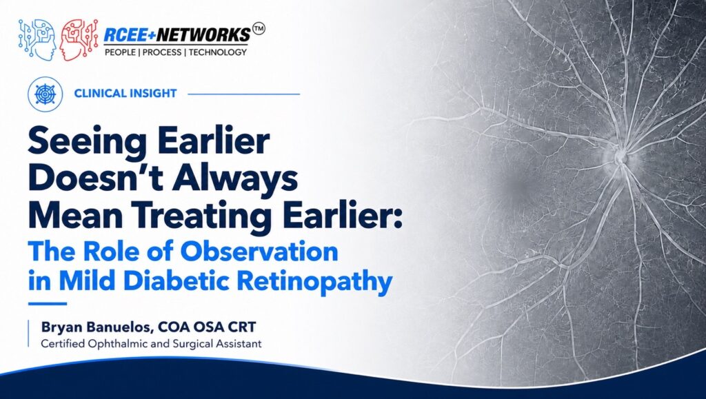
When disease burden is mild, vision remains stable, and no high-risk features are present, clinicians may recommend close follow-up rather than immediate anti-VEGF therapy, focal laser, or other intervention. Follow-up typically includes serial fundus photography, OCT- particularly when diabetic macular edema is suspected- visual acuity assessment, and comparison with prior imaging to identify subtle progression. Comparing current findings with historical retinal images often provides the most valuable clinical context. Side-by-side review helps determine whether microaneurysms, hemorrhages, or edema remain stable or represent meaningful progression that may alter management.
Avoiding unnecessary intervention may reduce treatment burden, procedural risks, patient anxiety, and healthcare costs while preserving excellent visual outcomes when careful surveillance is appropriate. At the same time, observation requires partnership. Patients who travel from rural areas where access to care is more challenging may benefit from earlier intervention simply because they are more likely to get lost to follow-up over time as I have seen in my clinical experience.
As retinal imaging continues to improve, clinicians will detect diabetic retinopathy at increasingly earlier stages. The greatest challenge is no longer simply identifying disease, but determining which patients require treatment today and which can be safely managed through careful surveillance. High-quality documentation and consistent longitudinal imaging remain essential to making that distinction. Reliable access to historical retinal images supports more confident clinical decision-making by allowing providers to compare disease progression over months or years rather than relying on a single examination.