Computational retinal image analysis uses AI and image processing to improve the early detection, diagnosis, and monitoring of retinal diseases. As the prevalence of vision-threatening conditions continues to rise worldwide, healthcare providers need faster and more accurate methods to evaluate retinal images. Eye diseases such as diabetic retinopathy, glaucoma, age-related macular degeneration (AMD), and hypertensive retinopathy often develop gradually and may not show noticeable symptoms in their early stages. Identifying these subtle retinal changes as early as possible is essential for preventing vision loss and improving treatment outcomes.
Key Takeaways
- Enhances retinal disease detection with AI and image processing.
- Supports the early diagnosis of major eye conditions.
- Improves screening efficiency and clinical decision-making.
- Advances teleophthalmology and personalized eye care.
Traditionally, retinal images have been examined manually by ophthalmologists, a process that requires considerable time and expertise. However, the growing demand for retinal screening has increased the need for automated and intelligent diagnostic solutions. Computational retinal image analysis addresses this challenge by analyzing retinal images efficiently, detecting abnormalities, and providing objective clinical insights that support ophthalmologists in making informed decisions. This article explores how computational retinal image analysis works, the technologies behind it, its clinical applications, key benefits, challenges, and future developments in modern ophthalmology.
What Is Computational Retinal Image Analysis?
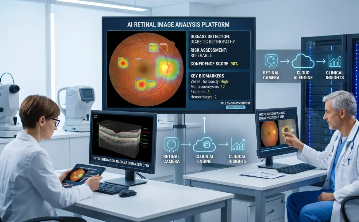
Computational retinal image analysis is the use of computer algorithms to examine retinal images and extract clinically relevant information. Instead of relying solely on manual observation, the technology processes digital images to identify anatomical structures, measure retinal features, and detect abnormalities that may indicate disease. The analysis can be performed on images captured through fundus cameras, optical coherence tomography (OCT), or other retinal imaging systems. By automatically evaluating these images, computational methods provide objective measurements and decision-support information that help eye care professionals make more informed clinical decisions. Rather than replacing ophthalmologists, computational retinal image analysis serves as an intelligent assistant that improves efficiency, supports accurate diagnosis, and enables large-scale retinal screening.
Also Read: Retinal Image Analysis Applications
How Computational Retinal Image Analysis Works
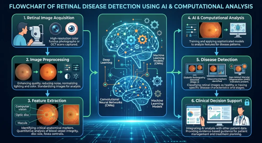
The analysis process follows a series of computational steps designed to transform raw retinal images into meaningful clinical insights.
1. Image Acquisition
The process begins with high-quality retinal images obtained using fundus photography, OCT, or other imaging technologies. Clear and well-focused images are essential because image quality directly influences the accuracy of subsequent analysis.
2. Image Preprocessing
Before analysis begins, the images undergo preprocessing to improve their quality. This stage may include noise reduction, brightness adjustment, contrast enhancement, and normalization. These improvements make retinal structures more visible and reduce variations caused by different imaging conditions
3. Feature Extraction and Segmentation
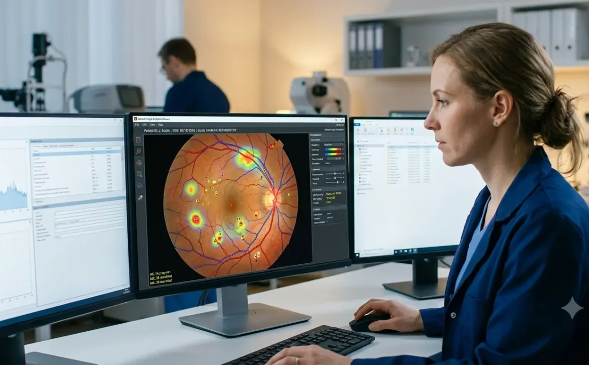
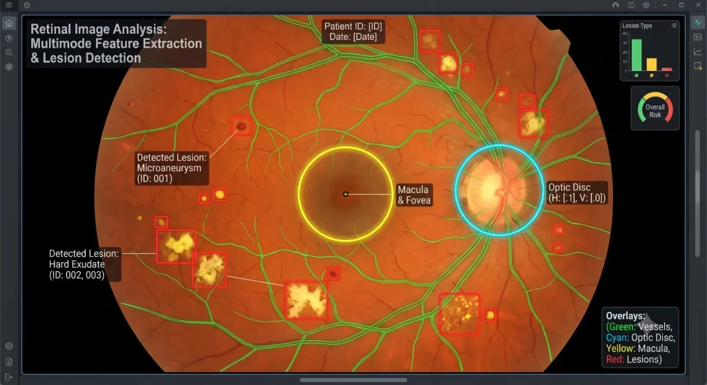
Advanced algorithms identify important retinal structures such as the optic disc, macula, blood vessels, and retinal layers. The system also detects abnormalities including microaneurysms, hemorrhages, exudates, drusen, and other pathological changes. Segmentation techniques isolate these structures so they can be measured accurately.
4. Disease Classification
After extracting relevant features, machine learning and deep learning models classify the retinal image based on the likelihood of disease. The results provide healthcare professionals with quantitative assessments that support clinical evaluation and patient management.
Key Computational Techniques Used
Several computational technologies work together to deliver accurate retinal image analysis.
- Image Processing improves image quality through Filtering, contrast enhancement, edge detection and morphological operations, allowing subtle retinal details to become more visible.
- Machine Learning uses carefully selected retinal features to recognize disease patterns. Traditional algorithms such as support vector machines and random forests remain useful in certain diagnostic applications.
- Deep Learning has transformed retinal image analysis by automatically learning complex visual patterns from large datasets. Convolutional Neural Networks (CNNs) and other deep learning architectures can identify retinal abnormalities with impressive accuracy while minimizing manual feature engineering.
- Computer Vision enables automated recognition of retinal structures, lesion detection, image registration, and quantitative measurements that assist clinicians during diagnosis and follow-up examinations.
Applications of Computational Retinal Image
- Analysis Computational retinal image analysis has become an important tool for detecting and monitoring numerous eye diseases. For diabetic retinopathy, algorithms identify microaneurysms, hemorrhages, hard exudates, and other early retinal changes that may indicate disease progression.
- In glaucoma, computational analysis evaluates the optic nerve head, cup-to-disc ratio, and retinal nerve fiber layer to support early diagnosis before significant vision loss occurs.
- For age-related macular degeneration (AMD), automated systems detect drusen, pigment abnormalities, and other macular changes that help clinicians assess disease severity and monitor progression.
- The technology also assists in identifying hypertensive retinopathy, retinal vein occlusion, and various vascular abnormalities. In addition, researchers are increasingly exploring retinal biomarkers that may help estimate cardiovascular and neurological health risks, expanding the role of retinal imaging beyond ophthalmology.
Also Read: Automated Retinal Image Analysis
Benefits of Computational Retinal Image Analysis
The growing adoption of computational retinal image analysis offers several important advantages for healthcare providers and patients.
- Supports earlier detection of retinal diseases before symptoms become severe.
- Improves diagnostic consistency by reducing observer variability.
- Processes large numbers of retinal images efficiently during screening programs.
- Provides objective measurements that support clinical decision-making.
- Reduces the workload associated with manual image interpretation.
- Enables continuous monitoring of disease progression over time.
- Supports teleophthalmology and remote retinal screening in underserved areas.
These benefits contribute to more efficient eye care services while helping clinicians deliver timely and evidence-based treatment.
Challenges and Future Trends
Despite its rapid progress, computational retinal image analysis still faces several challenges. Image quality can vary depending on the imaging device and patient cooperation, which may affect algorithm performance. Developing reliable AI models also requires large, diverse, and accurately annotated datasets to minimize bias and improve generalizability. In addition, healthcare providers must address issues related to data privacy, regulatory compliance, and integration with existing clinical workflows.
Looking ahead, advances in explainable AI, foundation models, federated learning, and multimodal imaging are expected to further improve diagnostic accuracy and transparency. Combined with cloud computing and teleophthalmology, these innovations will make advanced retinal analysis more accessible, particularly in regions with limited access to specialist eye care.
Also Read: Conditions Predicted by Retinal Image Analysis
Conclusion
Computational retinal image analysis is transforming modern ophthalmology by combining image processing, artificial intelligence, and computer vision to support faster and more consistent retinal evaluations. From detecting diabetic retinopathy and glaucoma to monitoring age-related macular degeneration, this technology enables earlier diagnosis and more informed clinical decisions. Although expert interpretation remains essential, computational tools continue to enhance efficiency, improve screening programs, and expand access to high-quality eye care. As AI technologies continue to evolve, computational retinal image analysis is expected to play an increasingly important role in protecting vision and supporting personalized healthcare.
Frequently Asked Questions
1. What is computational retinal image analysis?
Computational retinal image analysis uses computer algorithms, image processing, and AI to examine retinal images, identify abnormalities, and support ophthalmologists in diagnosing eye diseases.
2. Which eye diseases can computational retinal image analysis detect?
It can help detect diabetic retinopathy, glaucoma, age-related macular degeneration (AMD), hypertensive retinopathy, retinal vein occlusion, and other retinal disorders.
3. How does AI improve computational retinal image analysis?
AI automatically recognizes disease patterns, analyzes complex retinal features, and provides accurate decision-support information that enhances diagnostic efficiency.
4. Can computational retinal image analysis replace ophthalmologists?
No. It is designed to assist ophthalmologists by providing automated analysis and objective measurements, while the final diagnosis and treatment decisions remain the responsibility of qualified