
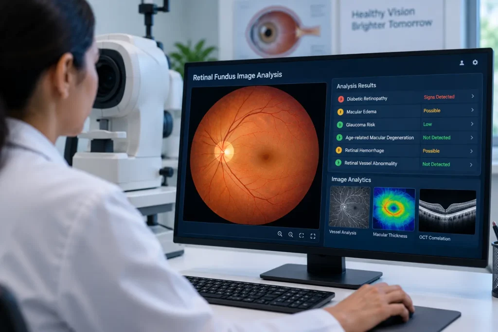
Retinal fundus image analysis detects eye diseases early by identifying subtle retinal changes, enabling faster diagnosis and timely treatment.Vision-threatening conditions often develop gradually, with few or no symptoms during their early stages, making routine retinal examinations essential for preserving long-term eye health. By analyzing detailed photographs of the retina, retinal fundus image analysis enables clinicians to assess important structures such as the optic disc, macula, and retinal blood vessels with greater accuracy.
Key Takeaways
- Retinal fundus image analysis
- Retinal fundus image analysis for diagnosis of glaucoma
- Retinal fundus image for glaucoma analysis dataset
- Retinal fundus images for glaucoma analysis the RIGA dataset
Combined with high-resolution fundus imaging, advanced image processing, and AI-assisted clinical decision support, this approach improves screening efficiency while supporting informed clinical decisions. Rather than replacing medical expertise, retinal fundus image analysis provides valuable insights that help ophthalmologists diagnose retinal diseases earlier and deliver timely, appropriate treatment.
What Is Retinal Fundus Image Analysis?
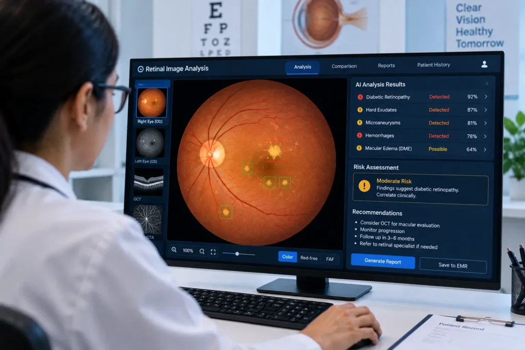
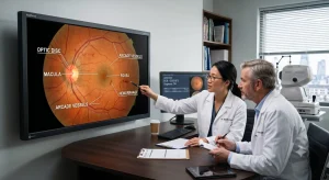
Retinal fundus image analysis refers to the examination of digital photographs of the back of the eye to identify healthy retinal structures and detect abnormalities. A fundus camera captures images of the optic disc, macula, retinal blood vessels, and surrounding tissues, providing valuable information about both ocular and systemic health.
Traditionally, specialists reviewed these images manually. Today, software-assisted analysis can automatically identify important retinal features, measure anatomical structures, and highlight suspicious findings for further evaluation. This approach increases consistency while helping clinicians review images more efficiently.
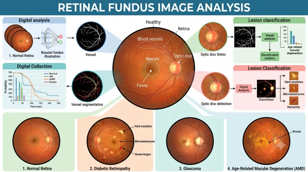
How Retinal Fundus Image Analysis Works
The process of retinal fundus image analysis involves several connected steps that transform a retinal photograph into clinically useful information.
- Image capture: A digital fundus camera records a clear image of the retina.
- Image enhancement: Brightness, contrast, and image quality are optimized while unwanted noise is minimized.
- Anatomical segmentation: The software identifies structures such as the optic disc, optic cup, retinal blood vessels, and macula.
- Feature measurement: Important clinical indicators, including the cup-to-disc ratio, vessel characteristics, and optic disc dimensions, are calculated.
- Clinical review: Ophthalmologists interpret the results alongside patient history and other diagnostic findings before making treatment decisions.
Also read: Computational Retinal Image Analysis
Applications of Retinal Fundus Image Analysis
The use of retinal fundus image extends across many areas of ophthalmology because a single retinal image can reveal signs of multiple diseases.
Common clinical applications include:
- Detecting diabetic retinopathy
- Supporting glaucoma assessment
- Monitoring age-related macular degeneration
- Evaluating hypertensive retinopathy
- Identifying retinal vascular disorders
- Assessing optic nerve health
Beyond hospitals, this technology is increasingly used in teleophthalmology, mobile eye screening units, and community healthcare programs where rapid evaluation of large numbers of retinal images is required.
Retinal Fundus Image Analysis for Diagnosis of Glaucoma
Early diagnosis is one of the most effective ways to slow glaucoma-related vision loss. Retinal fundus image analysis for diagnosis of glaucoma helps clinicians examine the optic nerve for subtle structural changes that may not be obvious during a routine eye examination.
Modern retinal fundus image analysis for diagnosis of glaucoma measures important biomarkers such as the cup-to-disc ratio, optic disc shape, neuroretinal rim thickness, and retinal nerve fiber layer appearance. When these measurements are combined with intraocular pressure testing, visual field assessment, and clinical examination, they provide a more complete picture of disease progression.
| Clinical Feature | Why It Matters |
| Cup-to-disc ratio | Indicates possible optic nerve damage |
| Optic disc morphology | Detects structural changes |
| Neuroretinal rim | Assesses nerve tissue loss |
| Retinal vessels | Supports optic nerve evaluation |
Also Read: Medical Software Company
Retinal Fundus Image for Glaucoma Analysis Dataset
Developing reliable diagnostic software requires carefully annotated retinal images. A retinal fundus image for glaucoma analysis dataset contains standardized retinal photographs with expert labels that identify the optic disc, optic cup, and glaucoma status.
Researchers use a retinal fundus image for glaucoma analysis dataset to train machine learning models, compare segmentation techniques, evaluate classification accuracy, and validate new image analysis methods. Public datasets also encourage collaboration by allowing researchers to benchmark their algorithms using the same reference images.
Retinal Fundus Images for Glaucoma Analysis: The RIGA Dataset
One of the most respected public resources in ophthalmic research is retinal fundus images for glaucoma analysis the RIGA dataset. The RIGA (Retinal Image Database for Optic Nerve Evaluation) dataset includes retinal photographs that have been independently annotated by multiple clinical experts.
Because retinal fundus images for glaucoma analysis the RIGA dataset contain multiple expert annotations, researchers can evaluate differences between human graders while improving automated optic disc and optic cup segmentation methods. This makes the dataset an important benchmark for validating glaucoma detection systems and advancing retinal imaging research.
Benefits and Future Outlook
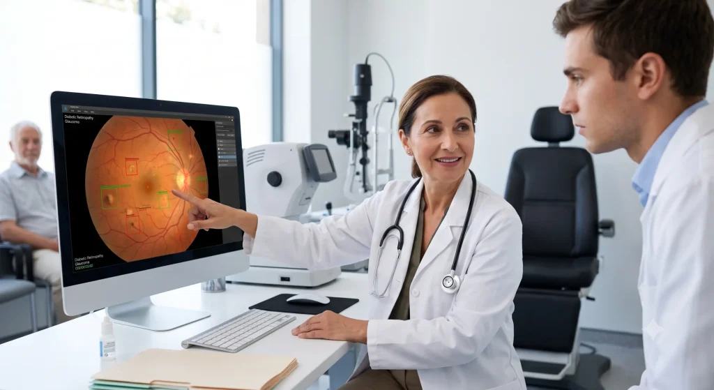
As retinal imaging technologies continue to improve, fundus image is becoming more accessible and more accurate. Automated image analysis helps reduce screening time, improves consistency, supports remote eye care services, and enables earlier detection of retinal diseases.
Future innovations are expected to deliver faster image processing, stronger clinical decision-support tools, and better integration with telemedicine platforms. These advances will allow healthcare providers to identify eye diseases earlier and expand access to quality eye care in both urban and rural communities.
Also Read: Medical Device Software Design
Conclusion
Retinal fundus image analysis has become an important part of modern ophthalmic practice by providing a detailed and objective assessment of retinal health. Its growing role in analysis for diagnosis of glaucoma supports earlier intervention and better long-term management of patients at risk of vision loss. At the same time, resources such as a fundus image for glaucoma analysis dataset and RIGA dataset continue to drive innovation in automated image analysis. As imaging technology advances, retinal fundus analysis will remain a valuable tool for improving the quality, accessibility, and efficiency of eye care.
Frequently Asked Questions
1. What is retinal fundus image analysis?
It is the process of evaluating retinal photographs to identify structural changes associated with eye diseases.
2. How is retinal fundus image analysis used for glaucoma?
It helps assess optic nerve features such as the cup-to-disc ratio and optic disc morphology to support glaucoma detection.
3. Why are glaucoma analysis datasets important?
They provide standardized, annotated retinal images that researchers use to train and evaluate automated diagnostic systems.
4. What makes the RIGA dataset valuable?
The RIGA dataset includes expert annotations from multiple graders, making it a trusted benchmark for glaucoma research.
5. Can automated retinal image analysis replace clinical diagnosis?
No. It serves as a clinical decision-support tool, while diagnosis and treatment decisions remain the responsibility of qualified ophthalmologists.
Pingback: Computational Retinal Image Analysis
Pingback: Retinal Image Analysis Applications
Pingback: AI Retinal Image Analysis Tools