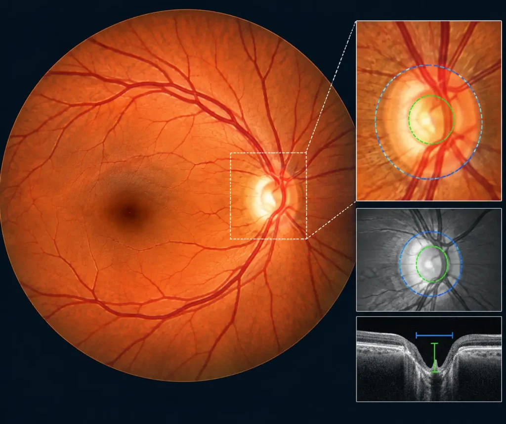
Glaucoma is a serious eye condition that can slowly damage the optic nerve. What makes it difficult to detect is that the early stages often do not cause noticeable symptoms. A person may feel that their eyesight is normal while changes are already taking place in the optic nerve. A Retinal Fundus Image Analysis for Diagnosis of Glaucoma can help researchers study these changes by providing retinal photographs that show the optic disc, optic cup, blood vessels, and surrounding retinal structures.
Key Takeaways
- Fundus datasets support glaucoma image-analysis research.
- Optic disc and cup annotations improve analysis accuracy.
- Image quality and clinical labels are essential.
- AI-powered tools can support glaucoma screening.
Also Read: AI-Powered Retina Image Analysis Market
This is why regular eye examinations and early screening matter. One useful tool in this process is retinal fundus image analysis. By examining photographs of the back of the eye, eye care professionals can look for structural changes around the optic nerve that may be linked to glaucoma.
Digital image analysis can also provide additional support during screening. When combined with clinical examination and Retinal Fundus Image Analysis for Diagnosis of Glaucoma , it can help doctors assess suspicious findings more efficiently.
What Is Retinal Fundus Image Analysis for Diagnosis of Glaucoma?
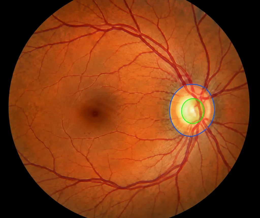
Retinal Fundus Image Analysis for Diagnosis of Glaucoma is an image of the inside back portion of the eye. It shows the retina, optic disc, optic cup, macula, and retinal blood vessels.
The clinician may look at the shape of the optic disc, the amount of cupping, the appearance of the nerve tissue around the cup, and differences between the two eyes.
Fundus image analysis may focus on several features, including:
· Optic disc shape and size
· Optic cup characteristics
· Cup-to-disc ratio
· Neuroretinal rim appearance
· Retinal nerve fiber layer visibility
· Differences between the right and left eyes
· Overall quality of the photograph
No single feature can confirm glaucoma.
How Can Fundus Images Help Identify Glaucoma?
Glaucoma damages nerve fibers that carry visual information from the retina to the brain. As this damage progresses, changes may become visible around the optic nerve head.
Examining the Optic Disc
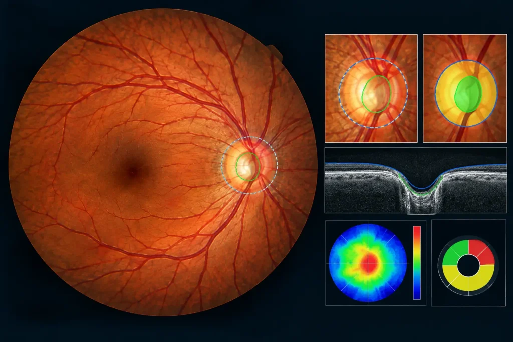
The optic disc is the area where the optic nerve exits the eye. During a fundus examination, doctors assess its shape, margins, color, and surrounding nerve tissue.
Changes in the appearance of the disc can provide clues that further glaucoma testing may be necessary for Retinal Fundus Image Analysis for Glaucoma Diagnosis
Looking at the Optic Cup
The optic cup is the central depression within the optic disc. In some people with glaucoma, progressive nerve loss causes the cup to become more pronounced.
However, a large optic cup is not automatically a sign of disease. Normal eye anatomy varies considerably from person to person, so the finding needs to be interpreted carefully.
Measuring the Cup-to-Disc Ratio
The cup-to-disc ratio (CDR) describes the size of the optic cup in relation to the optic disc.
A relatively large CDR or a substantial difference between the two eyes can attract clinical attention. It is not, however, a standalone diagnostic measurement.
Assessing the Neuroretinal Rim
Retinal Fundus Image Analysis for Diagnosis of Glaucoma the tissue surrounding the optic cup is called the neuroretinal rim. Glaucoma can cause this tissue to become thinner, sometimes in specific regions of the optic disc.
Examining the rim can therefore provide another piece of information when evaluating the optic nerve.
How AI-Powered Clinical Decision-Support Can Assist
Modern retinal imaging systems can use AI-powered clinical decision-support to examine fundus photographs and identify image patterns that may be associated with glaucoma.
A system may first determine whether an image is suitable for analysis. It can then locate the optic disc and optic cup, measure relevant features, and provide an assessment that can be considered by the clinician.
A simplified workflow is:
Fundus Photograph → Image Quality Check → Optic Disc Identification → Optic Cup Assessment → Feature Analysis → Clinical Review
The important point is that this technology provides support.
Key Features Examined in Retinal Fundus Image Analysis for Diagnosis of Glaucoma
| What It Tells the Clinician | |
| Optic disc | Provides information about the appearance of the optic nerve head |
| Optic cup | Helps identify changes in the central portion of the optic disc |
| Cup-to-disc ratio | Describes the relationship between the optic cup and disc |
| Neuroretinal rim | Helps assess the amount and distribution of nerve tissue |
| Inter-eye difference | Can reveal structural differences between the two eyes |
| Image quality | Determines whether the photograph is suitable for reliable assessment |
Where Can Retinal Image Analysis Be Used?
Retinal Fundus Image Analysis for Diagnosis of Glaucoma can be useful in several settings, particularly where large numbers of people need to be screened.
Potential applications include:
- Community eye screening programs
- Primary eye care centers
- Ophthalmology clinics
- Teleophthalmology services
- Mobile eye screening units
- Rural and underserved areas
- Long-term monitoring of retinal photographs
For remote screening, digital images can be captured locally and reviewed by specialists or analyzed with appropriate clinical decision-support systems.
Also Read:Retinal Fundus Image Analysis
Fundus Photography and OCT: Why Both Matter
Fundus photography and optical coherence tomography (OCT) provide different information.
A fundus photograph gives a direct visual record of the retina and optic nerve. It is useful for documenting changes in appearance over time.
OCT provides detailed cross-sectional images of retinal layers. It can also provide quantitative measurements, such as retinal nerve fiber layer thickness, which can be valuable when evaluating glaucoma.
Using both approaches can provide complementary information during glaucoma assessment.
Limitations to Keep in Mind
Retinal Fundus Image Analysis for Diagnosis of Glaucoma has an important role, but it has limitations. A photograph may be difficult to interpret if it is blurred, poorly illuminated, or affected by media opacity. Anatomical differences between individuals can also make image-based assessment more complicated.
Most importantly, glaucoma diagnosis does not depend on a single photograph.
Depending on the patient, a glaucoma evaluation may involve:
- Intraocular pressure measurement
- Visual field testing
- OCT imaging
- Optic nerve examination
- Gonioscopy
- Medical and family history
- Repeated examinations over time
These findings help the clinician distinguish glaucoma from normal variations in optic nerve appearance and other eye conditions.
Conclusion
Retinal Fundus Image Analysis for Diagnosis of Glaucoma is a useful part of modern eye care. By examining the optic disc, optic cup, neuroretinal rim, and other retinal features, clinicians can identify structural findings that may require closer investigation.
AI-powered clinical decision-support can assist with this process by examining digital retinal photographs and highlighting patterns of potential concern. Its value is greatest when it works alongside professional examination and established diagnostic tests.
For patients, the main benefit is simple: suspicious changes can be identified earlier, giving eye care professionals an opportunity to investigate them before substantial vision loss occurs.
FAQs
1.Can a retinal fundus image confirm glaucoma?
Not by itself. A fundus photograph can reveal structural changes that raise suspicion, but diagnosis usually requires a broader clinical assessment.
2.What does the cup-to-disc ratio mean?
It compares the size of the optic cup with the size of the optic disc. It is an important observation during optic nerve assessment, but a high ratio alone does not mean that someone has glaucoma.
3.Can AI-powered systems detect glaucoma from fundus photographs?
They can analyze retinal photographs for patterns associated with glaucoma and provide clinical decision-support. Their results should be interpreted by an appropriately qualified eye care professional.
4.Is a normal fundus photograph enough to rule out glaucoma?
No. Glaucoma can present in different ways, and some patients require additional tests to detect or evaluate the disease.
5.Is OCT better than fundus photography for glaucoma?
They serve different purposes. Fundus photography shows the visible appearance of the optic nerve and retina, while OCT provides detailed structural measurements. Both can contribute useful information during glaucoma evaluation.