
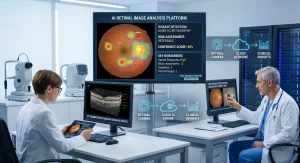
Artificial intelligence is reshaping healthcare, and ophthalmology is among the specialties experiencing significant advancements. AI retinal image analysis tools help eye care professionals detect retinal diseases faster and more accurately by analyzing high-resolution retinal images. Powered by advanced algorithms and AI-powered clinical decision-support, these tools assist clinicians in identifying subtle retinal abnormalities that might otherwise be difficult to detect during routine examinations.
Key Takeaways
- AI improves retinal disease detection.
- Supports clinical decision-making .
- Enhances screening efficiency .
- Expands access to eye care .
Rather than replacing ophthalmologists, AI retinal image analysis tools serve as valuable decision-support systems that improve diagnostic confidence, streamline clinical workflows, and support earlier intervention. As the demand for efficient retinal screening continues to grow, these technologies are becoming an important part of modern eye care.
What Are AI Retinal Image Analysis Tools?
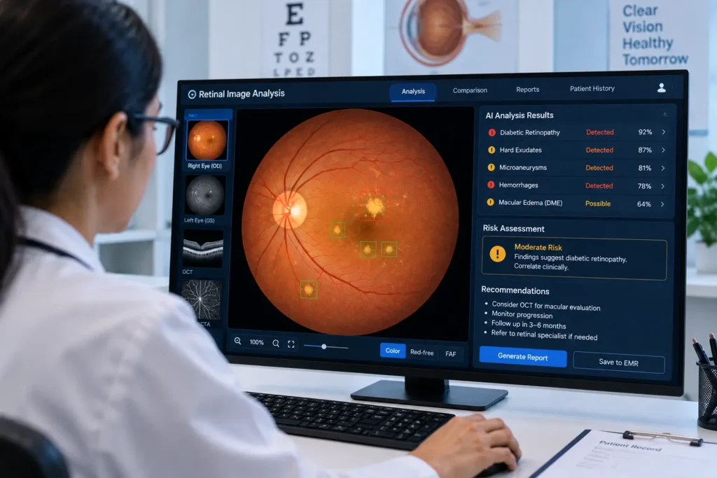
AI retinal image analysis tools are software platforms that analyze retinal photographs and optical coherence tomography (OCT) images using machine learning and deep learning techniques. They automatically evaluate retinal structures, detect abnormalities, and generate reports that help clinicians make informed decisions.
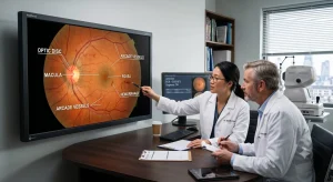
The process begins with capturing a high-quality retinal image using a fundus camera or OCT scanner. The software then enhances image quality, identifies important anatomical features such as the optic disc, macula, and retinal blood vessels, and analyzes them for signs of disease. Within seconds, the system provides severity scores, highlighted regions of concern, and clinical insights that support healthcare professionals during diagnosis.
Key Features of AI Retinal Image Analysis Tools
Modern AI retinal image analysis tools offer a wide range of features that improve both screening efficiency and diagnostic accuracy.
- Automated disease detection identifies retinal abnormalities with remarkable speed.
- Image quality assessment ensures only clinically usable images are analyzed.
- Retinal vessel analysis evaluates changes in blood vessels that may indicate ocular or systemic diseases.
- Optic disc and cup measurement supports glaucoma assessment by analyzing structural changes.
- Macular evaluation helps identify conditions affecting central vision.
- Disease progression monitoring compares current and previous scans to track changes over time.
- AI-powered clinical decision-support generates structured reports, probability scores, and visual heatmaps to assist clinicians without replacing their expertise.
These capabilities allow ophthalmologists to spend more time on patient care while improving consistency across examinations.
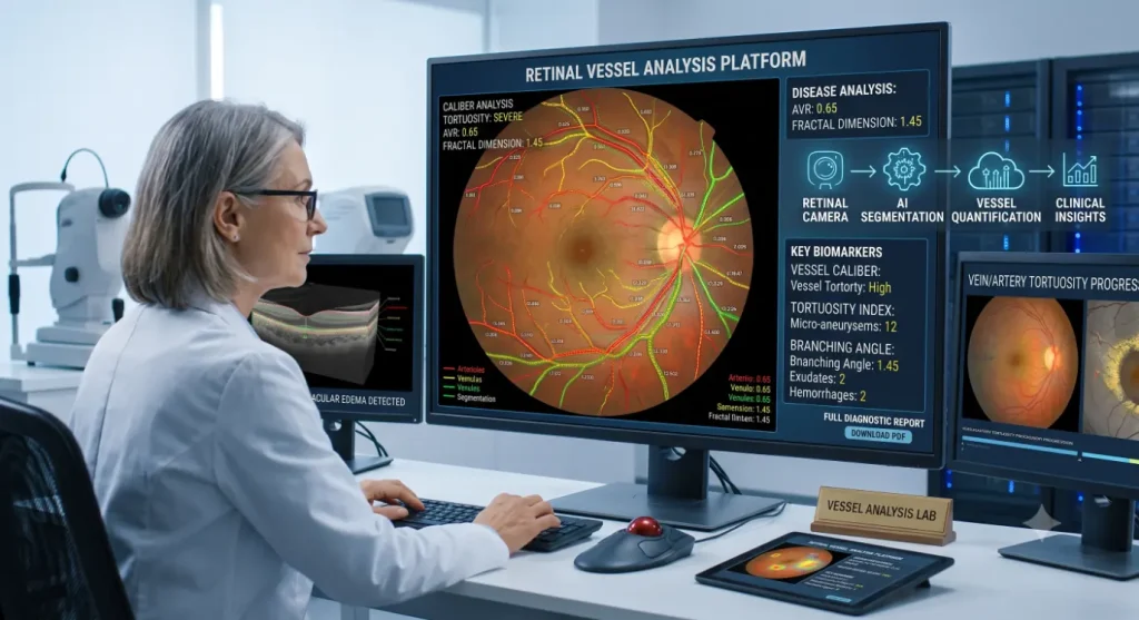
Eye Conditions Detected by AI Retinal Image Analysis Tools
AI retinal image analysis tools can assist in detecting several common retinal diseases during routine screening.
| Eye Condition | How the Tool Helps |
| Diabetic Retinopathy | Detects microaneurysms, hemorrhages, and exudates |
| Glaucoma | Evaluates optic nerve and cup-to-disc ratio |
| Age-Related Macular Degeneration | Identifies drusen and macular abnormalities |
| Diabetic Macular Edema | Detects retinal swelling and fluid accumulation |
| Hypertensive Retinopathy | Assesses vascular narrowing and hemorrhages |
| Retinal Vein Occlusion | Identifies retinal bleeding and vascular blockages |
In addition to eye diseases, researchers are exploring whether retinal biomarkers can help assess the risk of systemic conditions such as diabetes, cardiovascular disease, stroke, and chronic kidney disease. While these findings are promising, such applications are still evolving and should complement comprehensive medical evaluation rather than replace it.
Also Read:Conditions Predicted by Retinal Image Analysis
Benefits of AI Retinal Image Analysis Tools
- The adoption of AI retinal image analysis tools offers several advantages for healthcare providers and patients.One of the greatest benefits is early disease detection. Many retinal diseases develop gradually and may not produce noticeable symptoms until vision loss has already occurred. Automated image analysis helps identify subtle abnormalities at an earlier stage, allowing timely referral and treatment.
- These tools also improve diagnostic consistency by reducing variability between manual interpretations. Standardized analysis helps clinicians make more confident decisions, particularly during large-scale screening programs.
- Another important advantage is improved workflow efficiency. Automated analysis reduces the time required to review retinal images, enabling healthcare providers to examine more patients without compromising quality. This is especially valuable in busy ophthalmology clinics and diabetes screening programs.
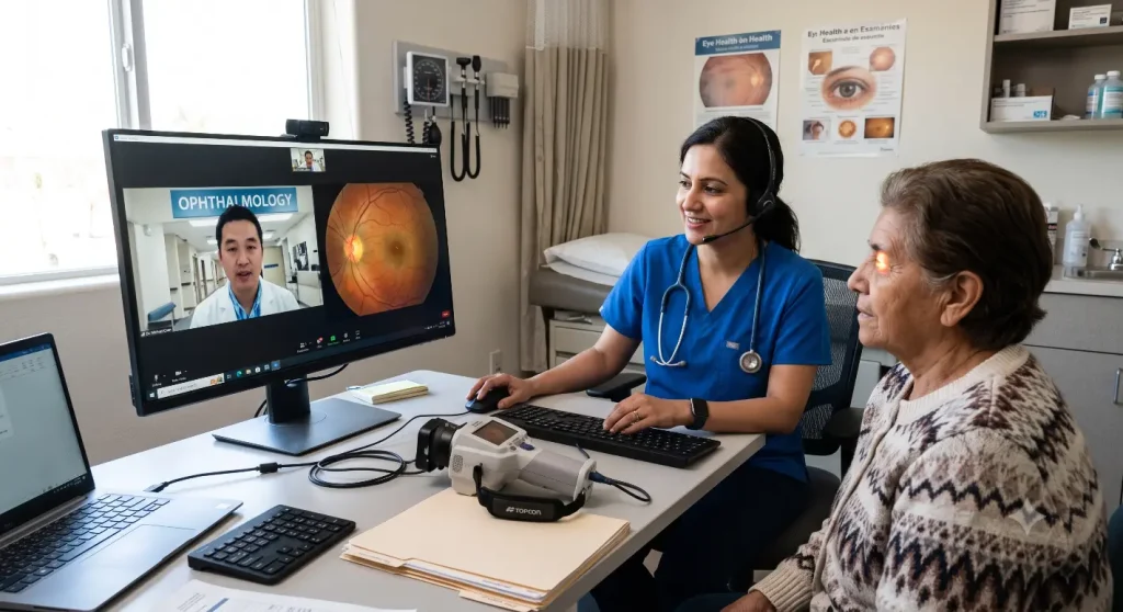
- AI retinal image analysis tools also support teleophthalmology, making retinal screening more accessible in rural and underserved communities where specialist eye care may be limited.
Limitations to Consider
Although AI retinal image analysis tools have transformed retinal screening, they are not without limitations. Poor-quality retinal images, media opacity, or inadequate lighting can affect analysis accuracy. In addition, algorithm performance depends on the diversity and quality of the data used during development.
False-positive and false-negative results remain possible, highlighting the importance of clinical oversight. AI-powered clinical decision-support should always assist—not replace—the judgment of qualified ophthalmologists. Regulatory approval, patient privacy, cybersecurity, and integration with electronic health record systems are also important considerations before implementing these technologies in clinical practice.
Future of AI Retinal Image Analysis Tools
The future of retinal imaging is moving toward more intelligent and integrated healthcare solutions. Emerging systems are combining fundus photographs, OCT images, and patient clinical information to provide more comprehensive assessments.
Advances in explainable AI are also making it easier for clinicians to understand how software reaches its conclusions, increasing trust and transparency. Portable retinal cameras, cloud-based platforms, and telemedicine integration are expected to expand access to retinal screening, particularly in remote areas.
As research continues, AI retinal image analysis tools may play an even greater role in personalized risk prediction, disease monitoring, and preventive eye care.
Also Read : Retinal Image Analysis
Conclusion
AI retinal image analysis tools are transforming ophthalmology by enabling faster, more accurate, and more consistent retinal screening. Through AI-powered clinical decision-support, these technologies assist healthcare professionals in detecting diseases such as diabetic retinopathy, glaucoma, and age-related macular degeneration at earlier stages, when treatment is often most effective.
While these tools cannot replace comprehensive clinical examination or the expertise of ophthalmologists, they significantly enhance diagnostic efficiency and patient care. As innovation continues, AI retinal image analysis tools will play an increasingly important role in improving eye health, expanding access to screening, and supporting better clinical outcomes worldwide.
Frequently Asked Questions
1. What are AI retinal image analysis tools?
They are software solutions that analyze retinal images using advanced algorithms to assist healthcare professionals in detecting eye diseases.
2. Can AI retinal image analysis tools replace ophthalmologists?
No. They function as AI-powered clinical decision-support systems that support, but do not replace, professional medical judgment.
3. Which eye diseases can these tools detect?
They can assist in detecting diabetic retinopathy, glaucoma, age-related macular degeneration, diabetic macular edema, hypertensive retinopathy, and retinal vein occlusion.
4. Are AI retinal image analysis tools useful for telemedicine?
Yes. They enable remote retinal screening and help expand access to eye care in underserved communities.
Also Read: Retinal Fundus Image Analysis
Pingback: Conditions Predicted by Retinal Image Analysis
Pingback: Retinal Image Analysis