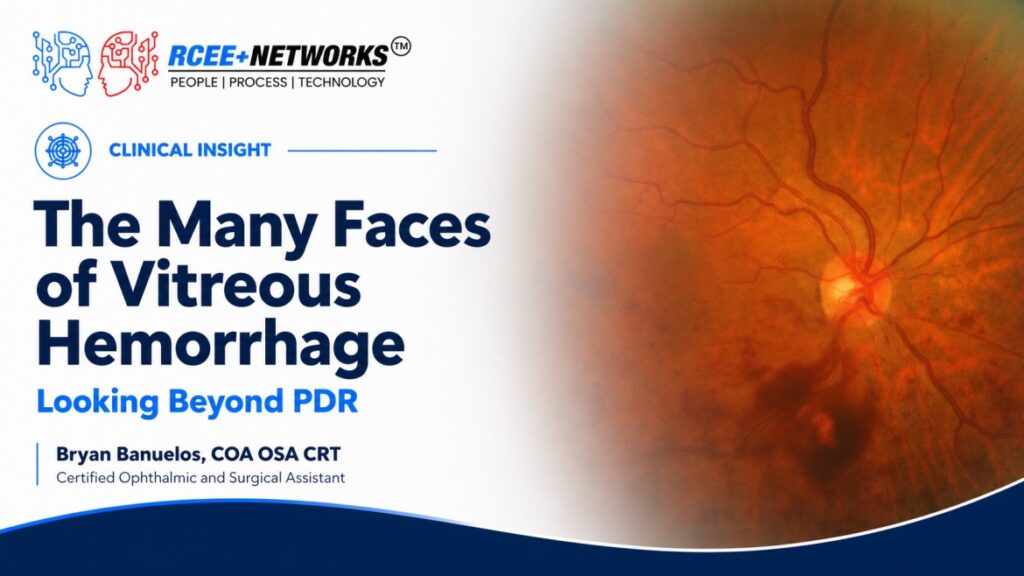
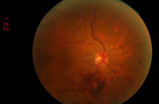
A recent case illustrates this point. Fundus examination revealed scattered microaneurysms and dot-blot hemorrhages consistent with diabetic retinopathy. However, the retinal hemorrhages were noticeably larger and predominantly concentrated in the inferior hemisphere. Additionally, the retinal veins appeared dilated and tortuous, raising suspicion for an inferior hemiretinal vein occlusion (HRVO). A small vitreous hemorrhage was also present inferior to the optic disc, raising concern that the patient may have progressed to PDR.
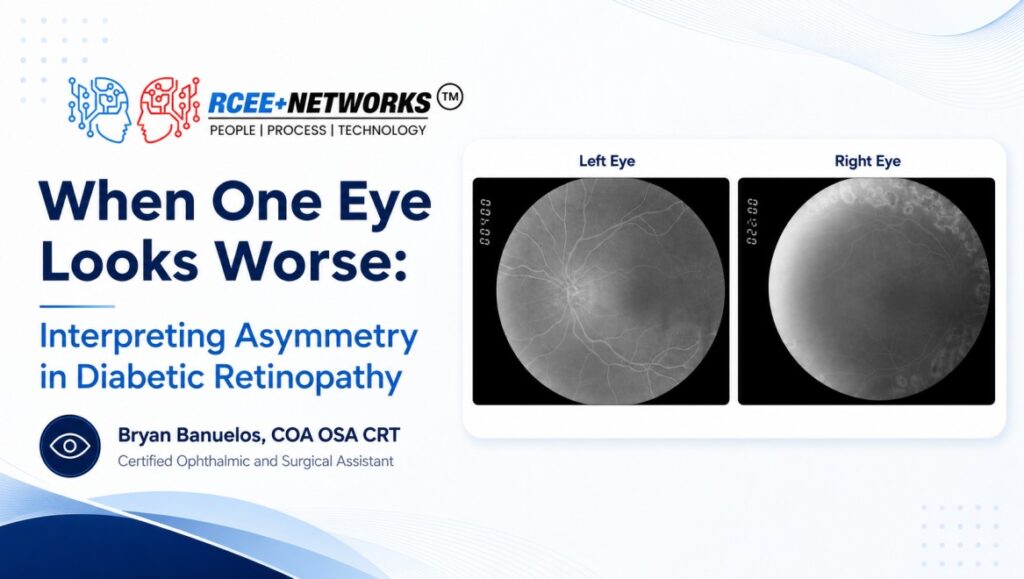
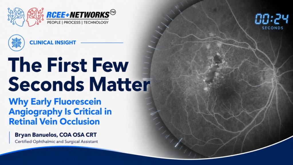
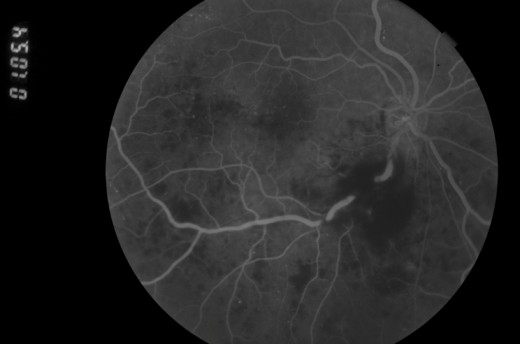
Fluorescein angiography (FA) provided the key to solving the case. Although the diabetic retinopathy and inferior HRVO were evident, FA showed no angiographic evidence of retinal or disc neovascularization. Instead, the vitreous hemorrhage was ultimately attributed to an acute posterior vitreous detachment (PVD), likely resulting from vitreoretinal traction and disruption of a superficial retinal vessel. In this patient, three separate pathologies coexisted: nonproliferative diabetic retinopathy, an inferior HRVO, and a symptomatic PVD.
This case serves as an important reminder that vitreous hemorrhage has a broad differential diagnosis. Vitreous hemorrhage may result from neovascularization associated with diabetic retinopathy or retinal vein occlusion, but it can also accompany an acute PVD without neovascular tissue. Distinguishing among these etiologies is critical because management strategies differ substantially.
PDR-associated neovascularization may require panretinal photocoagulation, intravitreal anti-VEGF therapy, or both. By contrast, a PVD-related hemorrhage may be managed with observation and close follow-up once a careful dilated peripheral retinal examination—and B-scan ultrasonography when visualization is limited—has excluded a retinal tear or detachment. Similarly, eyes with retinal vein occlusion require careful monitoring for the later development of retinal ischemia and secondary neovascularization.
For retina specialists and comprehensive eye care providers alike, the takeaway is simple: do not allow the patient’s medical history to prematurely narrow the differential diagnosis. A diabetic patient with a vitreous hemorrhage does not automatically have proliferative diabetic retinopathy. Careful evaluation of the hemorrhage pattern, retinal vasculature, posterior hyaloid status, and angiographic findings may reveal multiple coexisting pathologies. Recognizing each component is essential to reaching the correct diagnosis and selecting the appropriate management plan.
References:
American Academy of Ophthalmology. “Vitreous Hemorrhage.” EyeWiki. Accessed July 24, 2026.