Timely access to subspecialty care can significantly impact outcomes, particularly for patients with ocular tumors. We recently cared for a veteran whose case highlights how geographic barriers can delay treatment, even when the diagnosis is recognized early.
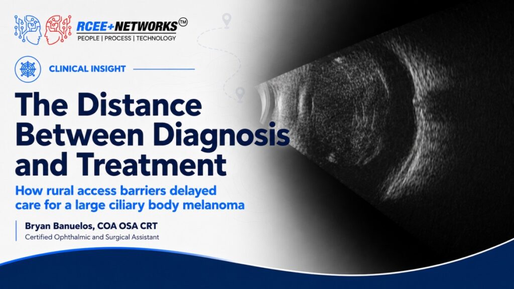
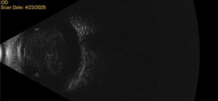
The patient was initially referred for evaluation of a suspected posterior segment mass. Examination revealed a shallow anterior chamber, a dense 4+ nuclear sclerotic cataract, and nasal posterior synechiae. Because the posterior segment could not be visualized, a B-scan ultrasonography was performed, revealing a large mass abutting the posterior lens with high suspicion for a ciliary body melanoma. An urgent referral to ocular oncology was made, and three weeks later enucleation was recommended, as the tumor was too large for plaque brachytherapy. Metastatic workup including chest and abdominal imaging, liver ultrasound, and hepatic function testing which was all negative.
Unfortunately, the patient was lost to follow-up.
One year later, he returned to our clinic. The tumor had enlarged considerably, secondary angle closure had progressed, and a prominent episcleral sentinel vessel was now present; an ominous clinical sign often associated with ciliary body melanoma. When asked what had happened, the patient explained that he lived in a rural community and simply could not find reliable transportation to the ocular oncologist, located five hours away. Although our retina clinic was only about an hour from his home, access to the nearest ocular oncology center proved to be a major barrier.
After repeating a metastatic workup which fortunately remained negative, the decision was made to proceed with enucleation locally. The surgery was uncomplicated.
Final pathology classified the tumor as Gene Expression Profile (GEP) Class 2, PRAME-negative. GEP testing has become an important prognostic tool in uveal melanoma. Class 1 tumors carry a relatively low metastatic risk, while Class 2 tumors have a substantially higher likelihood of systemic spread due to a more aggressive molecular signature. PRAME (Preferentially Expressed Antigen in Melanoma) further refines this risk. PRAME positivity is associated with an increased risk of earlier metastasis, whereas PRAME-negative tumors generally demonstrate a longer metastasis-free interval despite a Class 2 designation. Although this patient’s Class 2 result still warrants lifelong systemic surveillance, the negative PRAME status offers a somewhat more favorable prognosis than if both high-risk markers had been present.
This case is a reminder that barriers to care extend beyond insurance coverage or diagnosis. For many patients in rural America, the scarcity of ocular oncologists means hours of travel, dependence on family or transportation services, and difficult decisions that can delay sight- and life-saving treatment. As ophthalmologists, recognizing these challenges is just as important as recognizing the tumor itself.