Glaucoma is one of the leading causes of irreversible vision loss. One of the challenges is that it can develop slowly, often without obvious symptoms in the early stages. This is where retinal image analysis for detecting glaucoma can be useful. By examining images of the optic nerve and retinal structures, clinicians can identify changes that may need closer attention. Modern imaging tools can also provide measurements that help ophthalmologists track changes over time.
What Is Retinal Image Analysis for Detecting Glaucoma?
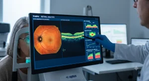
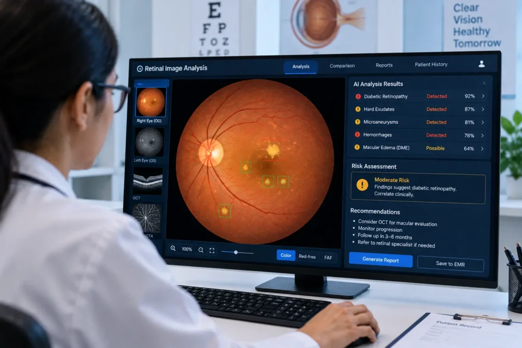
Retinal image analysis involves examining digital images of the retina and optic nerve to identify structural features associated with eye disease.
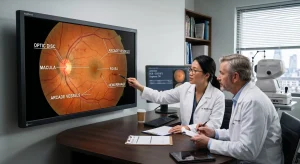
For glaucoma assessment, particular attention is paid to the optic nerve head, retinal nerve fiber layer, and ganglion cell layers.
Common imaging methods include:
- Fundus photography
- Optical coherence tomography (OCT)
- Optical coherence tomography angiography (OCTA)
- Digital retinal imaging
- Serial image comparison
Also Read:Medical Imaging Analysis Software
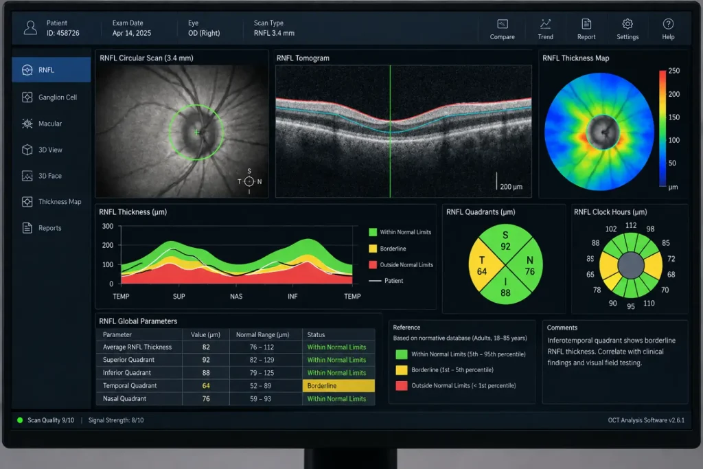
These technologies provide different types of information. A fundus photograph shows the appearance of the optic disc and surrounding retina, while OCT provides detailed cross-sectional measurements of retinal layers.
How Does Retinal Image Analysis Help Detect Glaucoma?
Glaucoma affects the optic nerve, and several structural changes can be associated with the disease. Retinal image analysis can help clinicians examine these changes more closely.
1. Optic Disc Analysis
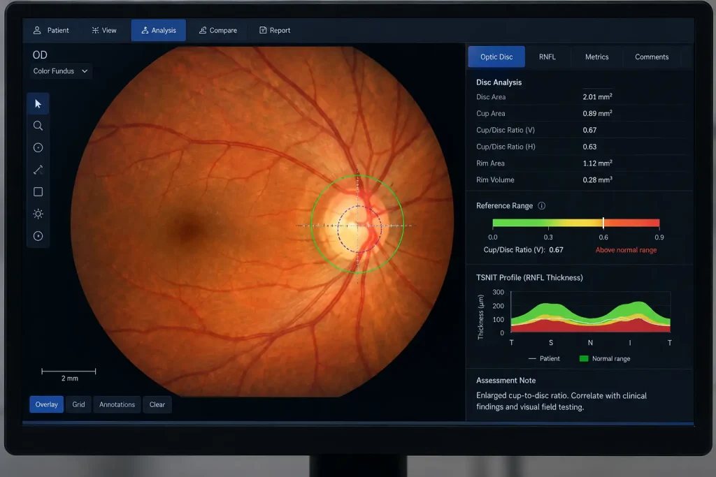
The optic disc is an important part of a glaucoma examination. Image analysis can help measure features such as the optic cup, optic disc area, and cup-to-disc ratio.
A larger or changing cup-to-disc ratio can be a clinical sign that requires further assessment. However, the measurement alone does not confirm glaucoma because normal optic nerve anatomy varies between individuals.
2. Retinal Nerve Fiber Layer Assessment
The retinal nerve fiber layer contains nerve fibers that carry visual information toward the brain. Glaucoma can cause these fibers to become thinner.
OCT can measure RNFL thickness around the optic nerve. Retinal image analysis can then help clinicians review these measurements and identify areas of possible thinning.
3. Ganglion Cell Analysis
Glaucoma can also affect retinal ganglion cells. OCT-based analysis can measure the thickness of specific layers around the macula.
Reviewing ganglion cell measurements together with RNFL findings can provide additional information when evaluating possible glaucomatous damage.
4. Comparing Images Over Time
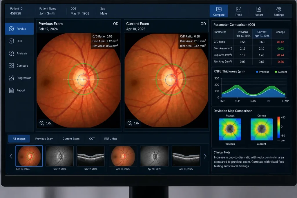
A single retinal image provides information about one point in time. Comparing images and OCT scans from different visits can be more useful for monitoring structural changes.
For example:
Previous image → Current image → Measurement comparison → Clinical assessment
This type of longitudinal review can help an ophthalmologist determine whether changes appear stable or require further investigation.
Also Read:Eyecare Software:Supports Eye Care Practices
Role of AI-Powered Clinical Decision-Support to Retinal Image Analysis for Detecting Glaucoma
An AI-powered clinical decision-support system can assist with the analysis of retinal images by identifying patterns or measurements that may be relevant to glaucoma assessment.
Depending on the technology, it may help with:
- Optic disc and cup analysis
- RNFL assessment
- Retinal layer measurements
- Image quality checks
- Identification of images that may need clinical review
- Comparison of imaging data from different visits
The system should be viewed as a support tool rather than a replacement for an ophthalmologist. A glaucoma diagnosis normally considers several factors, including eye pressure, optic nerve appearance, visual field results, medical history, and other examination findings.
Retinal Imaging Methods Used in Glaucoma Assessment
| Imaging method | Information it can provide |
| Fundus photography | Appearance of the optic disc and retina |
| OCT | RNFL and retinal layer measurements |
| OCTA | Information about retinal and optic nerve blood vessels |
| Digital image analysis | Quantitative measurements and image assessment |
| Serial imaging | Comparison of structural changes over time |
Benefits of Retinal Image Analysis
Retinal Image Analysis for Detecting Glaucoma screening and monitoring in several ways:
- Provides objective measurements of retinal structures
- Helps clinicians review optic nerve changes
- Supports comparison between previous and current scans
- Can assist with screening large patient populations
- May support teleophthalmology and remote image review
- Provides additional information alongside traditional glaucoma tests
Limitations to Consider
Retinal image analysis is not perfect. Poor image quality, cataracts, small pupils, unusual optic nerve anatomy, and other eye conditions can affect the results.
Glaucoma also cannot always be identified from structural imaging alone. Some patients may have visual field changes without obvious imaging abnormalities, while others may have unusual optic nerve structures with1.out having glaucoma.
For this reason, retinal imaging should be interpreted alongside other clinical information.
Frequently Asked Questions
1.Can Retinal Image Analysis for Detecting Glaucoma?
It can identify structural features that may be associated with glaucoma, but retinal imaging should not be considered a standalone diagnosis. A qualified eye-care professional needs to interpret the findings alongside other tests.
2.Which imaging technique is commonly used for Retinal Image Analysis for Detecting Glaucoma?
Fundus photography and OCT are commonly used. OCT is particularly useful for measuring the retinal nerve fiber layer and other retinal structures.
3.Can retinal image analysis detect glaucoma early?
It may help identify early structural changes, but results vary depending on the patient, imaging quality, disease stage, and technology used.
4.Is AI-powered clinical decision-support necessary for glaucoma detection?
Not necessarily. It is an additional tool that can assist with image analysis and clinical review. Traditional examination and diagnostic testing remain important.
5.Can retinal images be monitored over time?
Yes. Comparing retinal photographs and OCT scans from different visits can help clinicians monitor structural changes and support long-term glaucoma management.