Retinal Image Analysis for Age-related macular degeneration (AMD) affects the macula, the central part of the retina responsible for sharp, detailed vision. It is more common with age and can gradually affect activities such as reading, recognizing faces, and driving.
Key Takeaways
- Retinal imaging helps identify early AMD-related retinal changes.
- OCT can reveal fluid, swelling, and structural abnormalities.
- Image comparison supports long-term AMD monitoring.
- AI-powered clinical decision-support assists professional image review.
Because early AMD may not cause obvious symptoms, retinal imaging plays an important role in finding changes before vision is significantly affected. Retinal image analysis for age-related macular degeneration helps eye care professionals examine retinal images, identify suspicious findings, and monitor changes over time.
What Is Retinal Image Analysis for Age-Related Macular Degeneration?
AMD mainly affects the macula and is generally classified into two forms:
- Dry AMD: This is the more common form. It may involve drusen, pigment changes, and, in advanced stages, geographic atrophy.
- Wet AMD: This occurs when abnormal blood vessels develop beneath or around the retina.
Regular eye examinations are important because AMD can progress differently from one person to another.
How Retinal Image Analysis Helps With Retinal Image Analysis for Age-Related Macular Degeneration
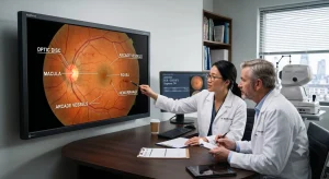
Retinal imaging provides clinicians with a detailed view of the retina. Depending on the imaging method, different features of AMD can be examined.
Detecting Drusen
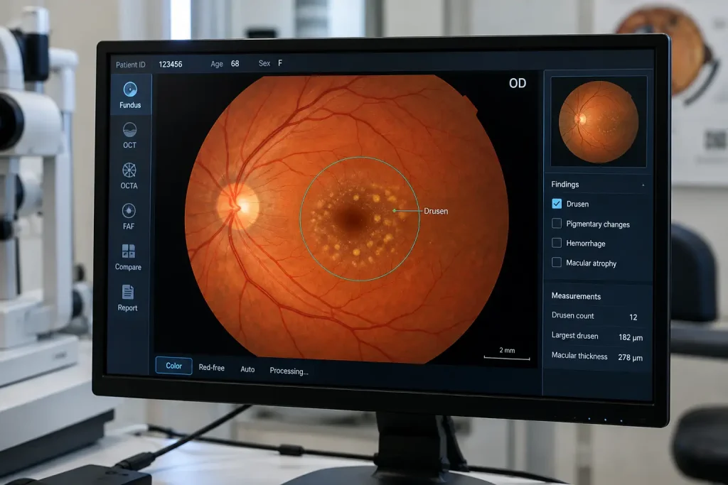
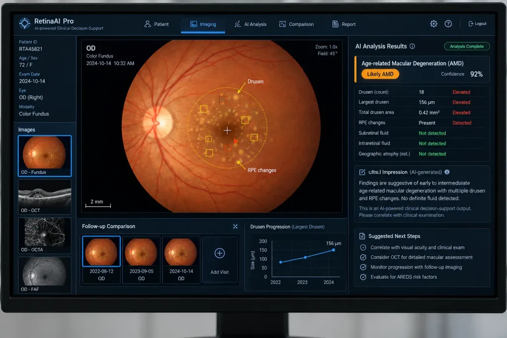
Drusen are deposits that can develop beneath the retina. Their size, number, and appearance can provide useful information when evaluating AMD. Fundus photography can make many of these changes visible.
Identifying Retinal Changes
Changes in pigmentation, retinal structure, or areas of tissue loss can provide additional information about disease progression.
Finding Fluid With OCT
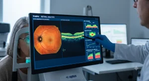
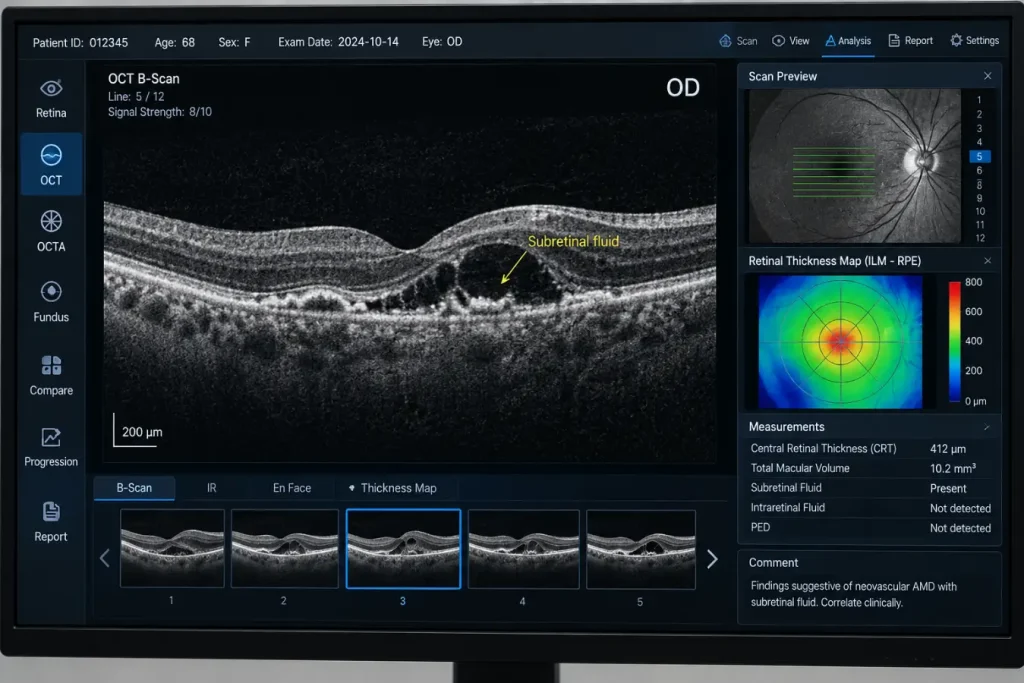
Optical coherence tomography, commonly known as OCT, creates cross-sectional images of the retina. It can help clinicians identify retinal fluid, swelling, changes in retinal layers, and other findings associated with wet AMD.
Monitoring Changes Over Time
A single retinal image provides a snapshot. Comparing images from different visits can give clinicians a better understanding of how retinal changes are developing.
Imaging Technologies Used in Retinal Image Analysis for Age-Related Macular Degeneration
Different imaging technologies provide different types of information.
| Imaging method | What it shows | Use in AMD |
| Fundus photography | A detailed photograph of the retina | Drusen, pigmentation, hemorrhages |
| OCT | Cross-sectional retinal structures | Fluid, swelling, atrophy, structural changes |
| OCTA | Blood-flow patterns without dye injection | Abnormal retinal or choroidal vessels |
| Fundus autofluorescence | Patterns related to retinal pigment changes | Assessment of retinal damage and atrophy |
Using more than one imaging method may be appropriate depending on the patient’s condition and the clinician’s assessment.
Role of AI-Powered Clinical Decision-Support
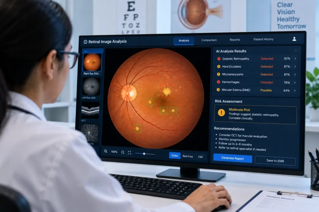
Retinal imaging software can now include AI-powered clinical decision-support to help clinicians review retinal images.
For AMD, this technology may support:
- Identification of possible drusen
- Detection of suspicious retinal abnormalities
- Assessment of fluid on OCT images
- Comparison of current and previous scans
- Screening of large numbers of retinal images
Why Image Comparison Matters
AMD is a condition that may change gradually. Keeping previous retinal photographs and OCT scans allows clinicians to compare current findings with earlier examinations.
This can help identify:
- New retinal abnormalities
- Changes in drusen
- Increasing or decreasing retinal fluid
- Progression of atrophic areas
- Changes following treatment
- Differences between the two eyes
A consistent record of retinal images can therefore be useful during routine follow-up.
Also Read: Medical Imaging Analysis Software
Retinal Image Analysis in Screening
Retinal image analysis can also be useful beyond individual eye clinics. Screening programs may collect large numbers of retinal images from older adults, community settings, mobile eye clinics, or teleophthalmology services.
Automated analysis can help flag images that require further assessment. This can be particularly useful when trained eye care professionals need to review a large volume of images.
However, image quality, patient characteristics, imaging devices, and software performance can affect results. Any suspicious finding should be assessed within the appropriate clinical context.
Key Benefits of Retinal Image Analysis for Age-Related Macular Degeneration
- Helps identify retinal changes associated with AMD
- Provides a visual record for future comparison
- Supports monitoring of disease progression
- Can assist clinicians when reviewing large numbers of images
- Works with imaging methods such as fundus photography and OCT
Frequently Asked Questions
1.Can retinal image analysis detect age-related macular degeneration?
Retinal imaging can identify findings associated with AMD, such as drusen, pigmentary changes, retinal fluid, and atrophy. A qualified eye care professional should interpret these findings and make the diagnosis.
2.Which imaging method is commonly used for Retinal Image Analysis for Age-Related Macular Degeneration ?
Fundus photography and OCT are commonly used. OCT is particularly useful for examining retinal structure and detecting fluid or other changes that may occur with wet AMD.
3.Can AI-powered clinical decision-support diagnose AMD?
It can assist with identifying image patterns associated with AMD, but it should be considered a clinical decision-support tool. Professional examination and interpretation remain important.
4.Why are previous retinal images useful?
Previous images provide a baseline for comparison. Looking at current and earlier images can help clinicians identify changes that may not be obvious from a single examination.
5.Is retinal image analysis useful for AMD screening?
Yes. It can support screening programs by helping identify retinal images that may need further clinical review.