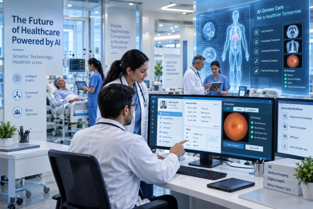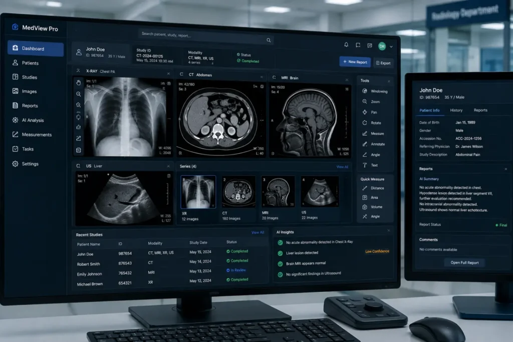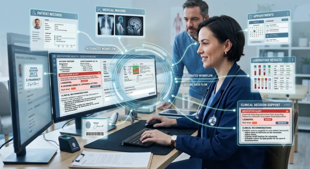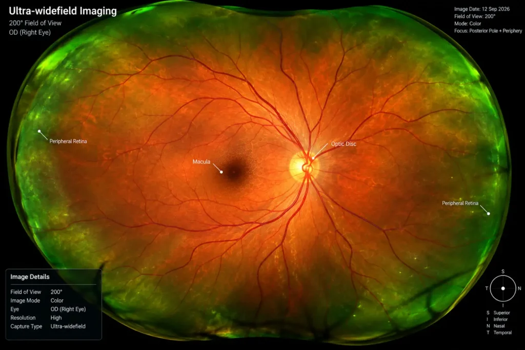
When people think about retinal imaging, they usually think about eye diseases. But the AI Retinal Imaging Full Body Health Analysis can offer information that goes beyond vision. It contains tiny blood vessels and nerve tissue that can reflect changes linked to conditions affecting the rest of the body.
Key Takeaways
- Retinal images can provide clues about more than eye health.
- AI-powered analysis can support clinicians in identifying health risks.
- Diabetes and hypertension can cause detectable retinal changes.
- Retinal AI supports clinical decisions but does not replace diagnosis.
With AI retinal imaging full body health analysis, retinal images can be examined for patterns that may be associated with diabetes, high blood pressure, cardiovascular risk, and other health conditions. The goal is not to diagnose every disease from an eye scan. Instead, AI-powered clinical decision-support can help clinicians notice potential warning signs and decide whether further testing may be needed.
What Is AI Retinal Imaging Full Body Health Analysis?
AI retinal imaging full body health analysis uses retinal photographs or other retinal scans with computer-based analysis to identify specific patterns or abnormalities.
A typical process looks like this:
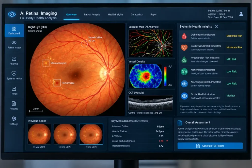
- A retinal image is captured using a fundus camera, OCT device, or another imaging system.
- The image is analyzed for relevant structural and vascular features.
- Potential abnormalities or risk indicators are highlighted.
- A healthcare professional reviews the findings.
- Additional tests are performed when clinically appropriate.
This distinction is important. Retinal image analysis can support healthcare decisions, but it should not be treated as a standalone diagnostic method for whole-body disease.
What Health Conditions Can Retinal Imaging Help Identify?
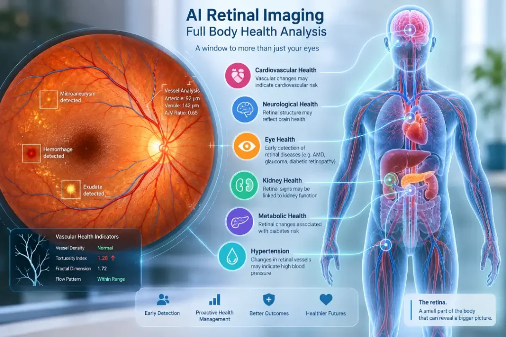
The retina is sensitive to changes in small blood vessels and neural structures. Some systemic conditions can affect these tissues, which is why researchers are exploring retinal imaging as a source of additional health information.
| Health condition | What retinal imaging may show |
| Diabetes | Changes associated with diabetic retinopathy |
| Hypertension | Abnormalities in retinal blood vessels |
| Cardiovascular risk | Vascular patterns associated with broader cardiovascular risk |
| Kidney disease | Retinal changes linked with vascular and metabolic conditions |
| Neurological conditions | Changes in retinal structures being studied as potential biomarkers |
| Eye diseases | Signs of glaucoma, AMD, diabetic macular edema, and other retinal conditions |
The strength of these associations varies. A retinal finding alone does not confirm that someone has a particular systemic disease.
Also Read: Medical Imaging Analysis Software
How AI-Powered Analysis Can Help
Reviewing retinal images requires training and careful attention to detail. AI-powered clinical decision-support can examine images systematically and highlight features that deserve closer clinical review.
Potential uses include:
- Detecting retinal abnormalities that may otherwise be missed
- Supporting diabetic eye screening
- Measuring retinal structures and vascular features
- Comparing current images with previous examinations
- Helping prioritize patients for additional assessment
- Supporting screening in primary-care and community settings
- Assisting large-scale retinal screening programs
For clinicians, the value is not simply automation. It is having another source of structured information to consider alongside the patient’s symptoms, medical history, examination, and test results.
Retinal Imaging Technologies Used for AI Retinal Imaging Full Body Health Analysis
Different imaging technologies provide different information.
Fundus photography captures images of the retina, optic disc, macula, and blood vessels. It is commonly used for retinal screening and documentation.
OCT, or optical coherence tomography, creates detailed cross-sectional images of retinal layers. It can reveal structural changes that may not be obvious in a standard photograph.
OCTA, or optical coherence tomography angiography, provides information about retinal and choroidal blood vessel networks.
These technologies can be combined with AI-powered clinical decision-support to analyze both visible and measurable retinal features.
Can an Eye Scan Really Assess Whole-Body Health?
This is where it is important to keep expectations realistic.
AI Retinal Imaging Full Body Health Analysis scan cannot replace a blood test, blood pressure measurement, medical examination, or specialist assessment. Not every disease produces detectable changes in the retina, and similar retinal findings can sometimes occur for different reasons.
For example, retinal vascular changes may be associated with hypertension, but the appropriate next step is still to measure blood pressure and carry out a proper clinical evaluation.
In other words, AI retinal imaging full body health analysis is better understood as a risk assessment and decision-support approach, not a complete health check from one image.
Also Read:Best Software for Reducing Administrative Burden in Medical Practices
Where Could It Be Useful?
The technology may have practical value in settings where early screening and access to healthcare are important.
Possible applications include:
- Community health programs
- Diabetes screening
- Mobile eye-care services
- Telemedicine
- Primary-care clinics
- Population-level screening
- Long-term disease monitoring
For people who may not regularly visit specialists, retinal imaging could provide an opportunity to identify findings that warrant further medical attention.
What Does the Future Look Like?
Research into retinal biomarkers is expanding, particularly around vascular, metabolic, and neurological conditions. As clinical evidence develops, retinal imaging may become a useful source of information for broader health risk assessment.
The most sensible approach is likely to combine retinal imaging, AI-powered clinical decision-support, clinician review, and appropriate confirmatory testing. This keeps the technology useful while avoiding the assumption that an algorithm can diagnose a person’s entire health profile from a retinal image.
FAQs
1.Can retinal imaging detect diseases outside the eye?
It can identify retinal patterns that may be associated with certain systemic conditions. However, these findings generally need clinical evaluation and, where appropriate, confirmatory testing.
2.Can AI Retinal Imaging Full Body Health Analysis detect diabetes?
Retinal imaging can identify signs of diabetic retinopathy. However, diabetes itself should be diagnosed using established clinical and laboratory tests.
3.Is AI retinal imaging a replacement for doctors?
No. AI-powered clinical decision-support is intended to assist healthcare professionals. Clinical judgment remains important when interpreting retinal findings.
4.What conditions are being studied through retinal imaging?
Research includes diabetes, hypertension, cardiovascular risk, kidney-related conditions, and neurological disorders, alongside established applications in eye diseases.
5.Is full-body health analysis from retinal images accurate?
Accuracy depends on the specific technology, dataset, intended use, image quality, and clinical validation. A retinal image should not be considered a complete diagnostic assessment of overall health.