Retinal Image Analysis Software opens a new door to the world. The field of ophthalmology is advancing rapidly. Technology has a crucial role in the diagnosis and management of various retinal diseases. Conditions such as retinopathy, glaucoma, and age-related macular degeneration affect millions of people worldwide, placing increasing pressure on healthcare providers to detect these diseases quickly and accurately. While conventional methods for reviewing retinal images are effective, they often require significant time, specialized expertise, and consistent evaluation.
Key Takeway:
- AI-powered clinical decision-support software assists earlier and more accurate detection of retinal diseases.
- Automated image analysis improves diagnostic consistency and clinical confidence.
- Streamlined workflows reduce manual effort and enhance patient care efficiency.
- Cloud-enabled platforms support scalable screening and remote ophthalmology services.
Modern retinal analysis software—powered by AI-powered clinical decision-support, machine learning (ML), and advanced image processing technologies—can assist physicians in various ways, such as detecting subtle retinal abnormalities, identifying potential signs of disease, and generating systematic reports within minutes. Rather than replacing healthcare professionals, these intelligent systems enhance clinical practice by providing reliable support and improving workflow efficiency.
Across the various settings—ranging from independent optometry clinics to multi-specialty hospitals and tele-ophthalmology programs—AI-powered clinical decision-support retinal imaging solutions are very helpful for eye care teams. it is very helpful for managing patient volumes while simultaneously maintaining high standards of care.
What Is Retinal Image Analysis Software?
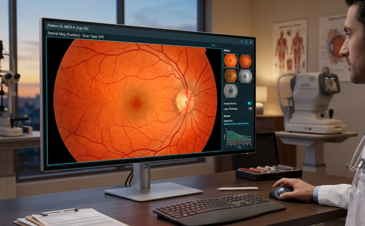
Retinal image analysis software is a digital platform that detects signs of disease by examining images of the retina—the light-sensitive tissue at the back of the eye. It monitors changes over time and assists in clinical diagnosis. Retinal image analysis software processes images captured by retinal imaging devices, such as fundus cameras or ultra-widefield imaging systems, to identify abnormalities that might not be immediately apparent through manual review alone, often utilizing advanced algorithms.
Unlike conventional image viewers, modern retinal analysis platforms integrate image enhancement, automated measurements, AI-powered clinical decision-support, and clinical reporting into a single workflow. This enables clinicians to review high-quality images more efficiently while simultaneously providing clinical decision-support-generated insights to support their assessments.
Why Retinal Image Analysis Matters More Than Ever?
The demand for retinal screening has grown significantly over the past decade. Rising rates of diabetes, an aging population, and increased awareness of preventive eye care have resulted in more patients requiring regular retinal examinations. While this is a positive development for public health, it also places greater demands on ophthalmologists and optometrists.
Manual image interpretation can become increasingly challenging as patient volumes rise. Each retinal image must be reviewed carefully, and even experienced clinicians may encounter subtle findings that require additional attention. AI-powered clinical decision-support retinal image analysis software helps address these challenges by streamlining image review and supporting faster, more consistent clinical assessments.
Earlier Detection of Vision-Threatening Diseases
Many retinal disorders develop gradually and may not produce noticeable symptoms in their early stages. By the time patients experience vision loss, irreversible damage may already have occurred.
Advanced image analysis software can identify subtle structural changes in retinal images, helping clinicians detect potential disease earlier and begin appropriate management before significant vision impairment develops.
Early detection is especially valuable for conditions such as:
- Diabetic retinopathy
- Age-related macular degeneration (AMD)
- Glaucoma
- Hypertensive retinopathy
- Retinal vascular disorders
Greater consistency in diagnosis.
Earlier intervention often leads to better long-term visual outcomes and can reduce the need for more complex treatments.
Variations in clinical interpretation among healthcare providers can naturally arise due to differences in experience, workload, and the complexity of a patient’s condition. AI-powered clinical decision-support-assisted analysis introduces a standardized approach; it evaluates retinal images using precise algorithms trained on vast datasets.
This additional analytical step helps reduce variability in results and—rather than replacing a clinician’s expertise—supports them in making more accurate, data-driven decisions.
Faster clinical workflows.
Time is a critical factor in eye care. Manually reviewing hundreds of retinal images can be extremely labor-intensive, particularly during community screening programs or in outpatient settings with high patient volumes.
Retinal image analysis software automates many routine tasks, including:
1. Image enhancement
2. Quality assessment
3. Detection of lesions or abnormalities
4. Quantitative measurements
5. Preliminary analysis
6. Report generation
Greater patient engagement.
By reducing the burden of administrative and repetitive tasks, clinicians can devote more time to patient care.
Visual explanations—such as images—are often easier for patients to understand than complex medical terminology. AI-powered retinal imaging platforms provide annotated images and structured reports that help clinicians explain examination results more clearly.
Enhanced care processes encourage patients to gain a better understanding of their condition, appreciate the importance of follow-up care, and participate more actively in treatment decisions.
Support for tele-ophthalmology services.
Access to specialized eye care remains limited in many remote and underserved areas. Using cloud-based retinal image analysis software, retinal images can be securely transmitted to specialists for remote review, making expert consultation accessible regardless of geographical location.
This capability facilitates large-scale screening programs and enables healthcare systems to expand the reach of timely eye care, thereby eliminating the need for every patient to physically visit a specialized medical center.
How Retinal image analysis software Works?
Although AI-powered clinical decision-support retinal analysis involves sophisticated technology, its workflow is designed to be straightforward and efficient for healthcare professionals.
Step 1: Image Acquisition
A Retinal image analysis softwareis captured using a compatible imaging device, such as a fundus camera, OCT system, or ultra-widefield retinal camera. High-quality images form the foundation for accurate analysis.
Step 2: Secure Retinal image analysis software Upload
The captured image is securely uploaded to the retinal image analysis platform, either through a cloud-based system or an on-premises deployment. Patient data is handled using appropriate security and privacy measures in accordance with applicable regulations.
Step 3: Image Quality Assessment
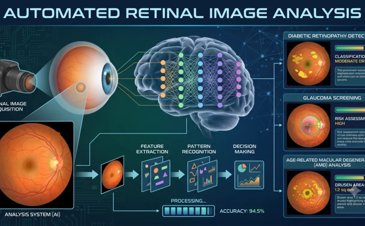
Before analysis begins, the software evaluates image quality using 5 key quality parameters: focus, brightness, contrast, field of view, and image clarity. Each retinal image is automatically assessed within 2–5 seconds to determine whether it is suitable for reliable interpretation. If the image fails to meet the predefined quality threshold, the system alerts the clinician and may recommend retaking the photograph. This automated quality check helps ensure that only high-quality retinal images proceed to AI analysis, improving the reliability of subsequent clinical assessments.
Step 4: AI-Powered Analysis
Advanced deep learning algorithms examine the retinal image to identify clinically relevant features, including microaneurysms, hemorrhages, exudates, optic disc changes, and other retinal abnormalities. The software compares detected patterns against trained models to estimate the likelihood of specific conditions.
Step 5: Clinical Report Generation
Once analysis is complete, the platform generates a structured report that summarizes key findings, highlights areas of concern, and provides visual annotations to support clinical review.
Step 6: Clinician Review and Decision
The eye care professional reviews both the original retinal image and the clinical decision-support-generated findings before making the final clinical assessment. The software serves as a decision-support tool, helping clinicians work more efficiently while maintaining full control over diagnosis and patient management.
Diseases Detected with Retinal Image Analysis Software
The retina provides valuable insights into eye health, often revealing signs of disease before symptoms appear. Powered by AI-powered clinical decision-support, retinal image analysis software helps clinicians identify abnormalities quickly, supporting earlier diagnosis and more informed clinical decisions.
Diabetic Retinopathy
Diabetic retinopathy is a leading cause of vision loss among people with diabetes. AI-powered clinical decision-support software can detect early signs such as microaneurysms, hemorrhages, and exudates, helping clinicians intervene before permanent damage occurs.
Age-Related Macular Degeneration (AMD)
AMD affects central vision and becomes more common with age. Retinal image analysis software assists in identifying drusen, pigment changes, and other retinal abnormalities that may indicate disease progression.
Glaucoma
Although glaucoma diagnosis requires multiple clinical tests, retinal analysis software can evaluate optic disc features, such as the cup-to-disc ratio, to support early detection and ongoing monitoring.
Hypertensive Retinopathy
High blood pressure can cause visible changes in the retinal blood vessels. AI-powered clinical decision-support-assisted analysis helps identify vascular abnormalities that may indicate hypertensive retinopathy and support timely medical evaluation.
Other Retinal Conditions
Depending on the platform, retinal image analysis software may also assist in detecting retinal vein occlusion, macular edema, optic nerve abnormalities, retinal lesions, and vascular changes. These insights
Why RetinaWiseAI™ Is Built for Modern Eye Care?
As eye care providers increasingly adopt digital technologies, an intelligent and efficient retinal imaging solution can bring about significant positive changes in both clinical workflows and patient care.
RetinaWiseAI™ is designed to support ophthalmologists, optometrists, hospitals, and screening programs through AI-powered clinical decision-support retinal image analysis; it simplifies the image review process and enhances workflow efficiency.
Its key features include:
• AI-powered clinical decision-support-assisted retinal image analysis
• Automatic detection of potential retinal abnormalities
• Streamlined clinical reporting
• Secure cloud-based access
• Compatibility with multiple retinal imaging devices
• Support for efficient screening and clinical workflows
By combining advanced clinical decision-support technology with a user-friendly platform, RetinaWiseAI™ enables healthcare professionals to review retinal images more efficiently while maintaining clinical oversight throughout the diagnostic process.
Also Read:Retinal Image Analysis – Early Detection of Eye Diseases.
How RCEENetworks Is Advancing AI-Powered Retinal Image Analysis
With the rising prevalence of retinal diseases, healthcare providers require smart technologies that can enhance operational efficiency while maintaining diagnostic quality. RCEENetworks is driving this transformation by developing AI-powered ophthalmology solutions that simplify retinal image analysis and facilitate improved clinical workflows.
Leveraging over 25 years of technological expertise, RCEENetworks combines AI-powered clinical decision-support, machine learning, cloud computing, and medical software development to create innovative solutions for ophthalmologists, optometrists, hospitals, and eye care networks.

Their flagship platform, ‘RetinaWiseAI™’, aims to assist clinicians in analyzing retinal images more efficiently, streamlining reporting processes, and rapidly detecting retinal diseases. The platform integrates with retinal imaging devices and cloud-based workflows, enabling healthcare teams to securely manage images while fostering collaboration and boosting operational efficiency.
In addition to RetinaWiseAI™, RCEENetworks offers services such as custom healthcare software development, IMACAM™ support, clinical decision-support integration, and technical consulting. Through these offerings, they help organizations modernize their ophthalmology workflows and prepare for the future of digital eye care.
By combining advanced technology with a deep understanding of clinical needs, RCEENetworks continues to empower healthcare providers to deliver faster, smarter, and more accessible retinal care.
Frequently Asked Questions (FAQs)
1. What is retinal image analysis software?
Retinal image analysis software is a digital solution that uses AI-powered clinical decision-support and image processing to assist clinicians in evaluating retinal images, identifying abnormalities, and supporting the diagnosis of eye diseases.
2. How does AI improve retinal image analysis?
AI can quickly analyze retinal images, detect patterns associated with common eye diseases, highlight areas of concern, and generate structured insights that help clinicians make informed decisions more efficiently.
3. Which eye diseases can retinal image analysis software help detect?
AI-powered clinical decision-support retinal image analysis can assist in identifying signs of diabetic retinopathy, age-related macular degeneration (AMD), glaucoma, hypertensive retinopathy, macular edema, and other retinal abnormalities.
4. Who can benefit from retinal image analysis software?
The software is designed for ophthalmologists, optometrists, eye clinics, hospitals, teleophthalmology programs, and healthcare organizations involved in retinal screening and diagnosis.
5. What features should you look for in retinal image analysis software?
Key features include AI-powered clinical decision-support-assisted analysis, image enhancement, automated reporting, cloud accessibility, EMR/EHR integration, multi-device compatibility, and secure data management.
6. How does RetinaWiseAI™ support eye care professionals?
RetinaWiseAI™ helps clinicians streamline retinal image analysis, improve workflow efficiency, generate structured reports, and support early identification of retinal abnormalities while maintaining clinical oversight.
7. Why choose RCEENetworks for ophthalmology technology solutions?
RCEENetworks combines over 25 years of technology expertise with AI, cloud computing, and medical software development to deliver innovative solutions that enhance retinal imaging workflows and support better patient care.
8. Is retinal image analysis software suitable for remote or multi-location healthcare practices?
Yes. Cloud-enabled retinal image analysis platforms allow authorized healthcare professionals to securely access images and collaborate across multiple clinics, hospitals, or remote screening programs, improving efficiency and continuity of care.
Learn More:https: https://www.rceenetworks.com/