The retina is a thin, light-sensitive layer located at the back of the eye. Often described as a “window to overall health,” it contains a rich network of blood vessels and nerve tissue that can reveal early signs of both eye diseases and certain systemic conditions.Discover conditions predicted by retinal image analysis using AI-powered clinical decision-support for early detection of eye and systemic diseases accurately.
Understanding which conditions can be predicted using retinal image analysis is becoming increasingly important as healthcare providers prioritize earlier disease detection, timely intervention, and improved patient outcomes. Although retinal image analysis does not replace a comprehensive clinical examination, it serves as a valuable clinical decision-support tool by enhancing diagnostic confidence, supporting risk assessment, and helping clinicians prioritize patients who require further evaluation and follow-up.
Key Takeways:-
- Early Disease Detection
- Glaucoma & AMD Screening
- AI-Powered Clinical Decision-Support in RetinaWiseAI
- Systemic Health Insights
What Is Retinal Image Analysis?
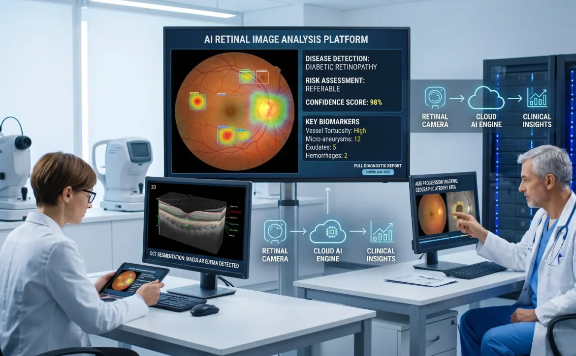
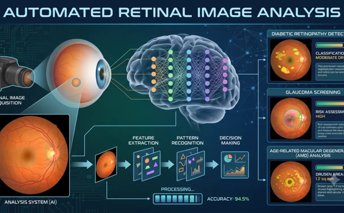
Retinal image analysis is the process of examining digital images of the retina captured through technologies such as fundus photography and Optical Coherence Tomography (OCT). High-resolution retinal images provide a clear and detailed view of the retina, optic nerve, macula, and retinal blood vessels, helping clinicians detect abnormalities and diagnose eye conditions more accurately. Modern image analysis systems use advanced algorithms and AI-powered clinical decision-support to assist eye care professionals in identifying patterns associated with various diseases. Rather than making an independent diagnosis, these systems highlight suspicious findings, measure structural changes, and support clinicians in making informed decisions of conditions predicted by retinal image analysis.
Why Is the Retina Important for Disease Detection?
The retina reflects changes occurring throughout the body. Because its blood vessels and nerve tissues can be viewed directly without invasive procedures, retinal imaging offers valuable insights into both ocular and systemic health. Changes in the retina may appear long before noticeable symptoms develop. Early identification of these changes allows healthcare professionals to recommend additional testing, monitor disease progression, and begin treatment before significant vision loss occurs.
Which conditions can be predicted using retinal image analysis ?
Retinal image analysis can assist clinicians in detecting and monitoring several important eye conditions.Diabetic retinopathy is one of the most common diseases of the retina and a leading cause of preventable blindness among diabetic patients. Therefore, timely detection and treatment are crucial. Elevated blood sugar levels can damage tiny retinal blood vessels, leading to microaneurysms, hemorrhages, fluid leakage, and abnormal blood vessel growth.Retinal image analysis helps detect early retinal changes, enabling timely referral and appropriate treatment before irreversible vision loss can occur. Regular retinal screening is an essential part of diabetes care, helping detect retinal changes early and enabling timely treatment to reduce the risk of vision loss.
Here are the 4 conditions predicted by retinal image analysis:
1.Retinal Image Analysis for detecting Glaucoma
Conditions Predicted by Retinal Image Analysis include glaucoma, which is detected by evaluating the optic nerve and retinal nerve fiber layer. Glaucoma gradually damages the optic nerve, often without noticeable symptoms until significant vision has already been lost. Image analysis tools can assess optic disc appearance, estimate the cup-to-disc ratio, and identify thinning of the retinal nerve fiber layer.
When combined with AI-powered clinical decision-support, these measurements help clinicians recognize individuals who may require additional glaucoma testing and long-term monitoring. Early detection plays a critical role in slowing disease progression and preserving vision. its also help for conditions predicted by retinal image analysis.
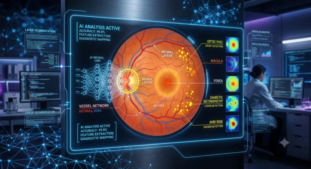
2.Retinal Image Analysis for Age-Related Macular Degeneration
Conditions predicted by retinal image analysis include age-related macular degeneration (AMD), making retinal image analysis an important application in modern ophthalmology. Age-related macular degeneration (AMD) is a progressive eye disease that affects the macula, the central part of the retina responsible for sharp, detailed vision needed for everyday activities such as reading, driving, and recognizing faces. Conditions predicted by retinal image analysis can identify early indicators such as drusen deposits, pigment changes, and structural abnormalities. OCT imaging further supports clinicians by visualizing fluid accumulation or retinal thinning associated with different forms of AMD.
Early identification allows appropriate monitoring and treatment strategies that may help preserve central vision. Diabetic macular edema (DME) develops when damaged retinal blood vessels leak fluid into the macula, causing swelling that can blur or distort central vision. Early detection is essential to help prevent long-term vision loss. Retinal imaging allows clinicians to measure retinal thickness and monitor fluid accumulation over time. Accurate assessment supports treatment planning and helps evaluate patient response during conditions predicted by retinal image analysis.
3.Hypertensive Retinopathy
High blood pressure can produce visible changes within the retinal blood vessels. Narrowed arteries, hemorrhages, cotton wool spots, and vascular abnormalities may indicate long-term hypertension. Retinal image analysis assists healthcare professionals in documenting these findings and assessing disease severity. In many cases, retinal changes also provide valuable information about overall cardiovascular health. its also focus on conditions predicted by retinal image analysis.
4. Retinal Vein and Artery Occlusions
Blockages affecting retinal veins or arteries can cause sudden vision loss and require urgent medical attention. Retinal imaging helps clinicians identify bleeding, swelling, interrupted blood flow, and other characteristic features. Rapid image analysis supports timely referral and appropriate clinical management.
Can Retinal Image Analysis Indicate Other Health Conditions?
Beyond eye diseases, researchers continue to explore whether retinal features can provide insights into broader health conditions. Research suggests that changes in the retinal blood vessels may be associated with systemic conditions such as diabetes, cardiovascular disease, chronic kidney disease, and an increased risk of stroke, highlighting the broader health insights that retinal image analysis can provide. Although these applications are promising, many remain under active investigation. At present, retinal image analysis should be viewed as a supportive clinical tool rather than a standalone method for predicting systemic disease.
Benefits of AI-Powered Clinical Decision-Support in Retinal Image Analysis
The integration of AI-powered clinical decision-support has significantly enhanced retinal image analysis by assisting clinicians with faster, more consistent image interpretation.
Key benefits include:
- Earlier identification of subtle retinal abnormalities.
- Improved screening efficiency in busy clinical settings.
- More standardized image evaluation across different healthcare providers.
- Better support for disease monitoring during follow-up visits.
- Enhanced access to retinal screening in remote and underserved communities through teleophthalmology.
Importantly, these systems are designed to support clinical judgment—not replace the expertise of ophthalmologists or optometrists. it is important for conditions predicted by retinal image analysis.
Limitations of Retinal Image Analysis
Despite its advantages, retinal image analysis has certain limitations. Poor image quality, media opacities such as cataracts, patient movement, or inadequate pupil dilation can reduce diagnostic accuracy. In addition, imaging findings must always be interpreted alongside a patient’s medical history, symptoms, and comprehensive eye examination. For this reason, retinal image analysis should be considered one component of a complete clinical assessment rather than a definitive diagnostic tool.
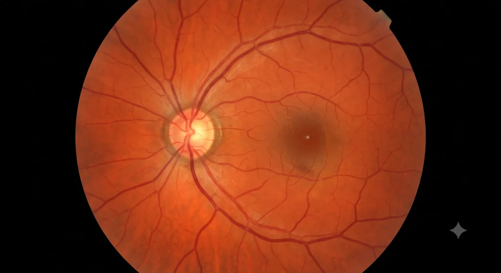
The range of conditions predicted by retinal image analysis continues to expand as imaging technologies and AI-powered clinical decision-support evolve. Today, retinal image analysis plays an important role in identifying diabetic retinopathy, glaucoma, age-related macular degeneration, diabetic macular edema, hypertensive retinopathy, and retinal vascular disorders. Ongoing research also suggests that retinal imaging may provide valuable insights into broader systemic health.
Regular retinal examinations are crucial if there is a family history of diabetes, high blood pressure, or eye diseases, or if there are age-related changes in vision. This helps in the early detection of serious eye conditions and ensures timely treatment.When combined with expert clinical evaluation, retinal image analysis supports earlier detection, more informed treatment decisions, and improved long-term eye health outcomes.Protect your sight before it’s at risk—because every clear view of life is worth preserving.
Also read:Retinal Image Analysis
Frequently Asked Questions
1. Which conditions predicted by retinal image analysis ?
Retinal image analysis helps detect a variety of vision-threatening eye diseases, including diabetic retinopathy, glaucoma, age-related macular degeneration (AMD), diabetic macular edema, hypertensive retinopathy, and retinal vascular occlusions, supporting earlier diagnosis and timely treatment.
2. How effective is retinal image analysis for detecting glaucoma?
Retinal image analysis evaluates the optic nerve and retinal nerve fiber layer, helping clinicians identify structural changes associated with glaucoma at earlier stages.
3. Can retinal image analysis detect age-related macular degeneration?
Yes. Retinal imaging can reveal drusen, pigment abnormalities, and other retinal changes that assist clinicians in detecting and monitoring AMD.
4. Does retinal image analysis replace an eye examination?
No. This is a clinical decision-support tool that complements a comprehensive eye examination performed by an eye specialist, but it is not a substitute for that examination.
Also Read: Retinal Image Analysis Software