Retinal diseases such as diabetic retinopathy, glaucoma, and age-related macular degeneration (AMD) are among the leading causes of preventable vision loss worldwide. Detecting these conditions at an early stage is essential for preserving vision and improving long-term patient outcomes. Automated retinal image analysis helps healthcare professionals evaluate retinal images faster and more consistently. It is supporting earlier eye disease screening. Instead of relying solely on manual interpretation, clinicians can now use advanced image-processing technologies supported by AI-powered clinical decision-support to identify retinal abnormalities and prioritize patients who may require further examination.
Key Takeways:
- Early Disease Detection
- AI-Powered Clinical Decision-Support
- Scalable Screening
- Future of Eye Care
Also known as automatic retinal image analysis, this technology analyzes high-resolution retinal images captured through fundus photography, optical coherence tomography (OCT), or other digital imaging devices. It examines retinal structures, detects disease-related patterns, and provides decision-support information that complements the expertise of ophthalmologists and optometrists. While these systems can significantly improve screening efficiency, they are designed to assist clinical judgment rather than replace comprehensive eye examinations.
What Is Automated Retinal Image Analysis?
Automated retinal image analysis is a computer-assisted process that evaluates digital retinal images using advanced image-processing algorithms and AI-powered clinical decision-support. The goal is to identify retinal features and patterns that may be associated with various eye diseases, allowing clinicians to review images more quickly and consistently.
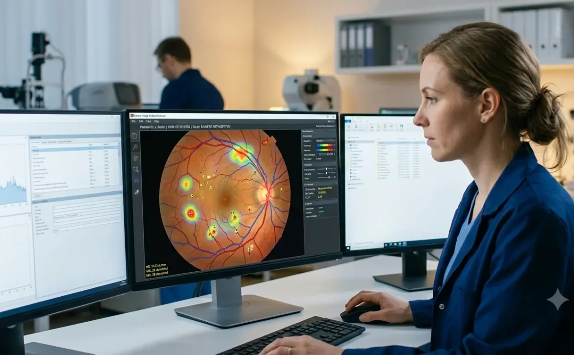
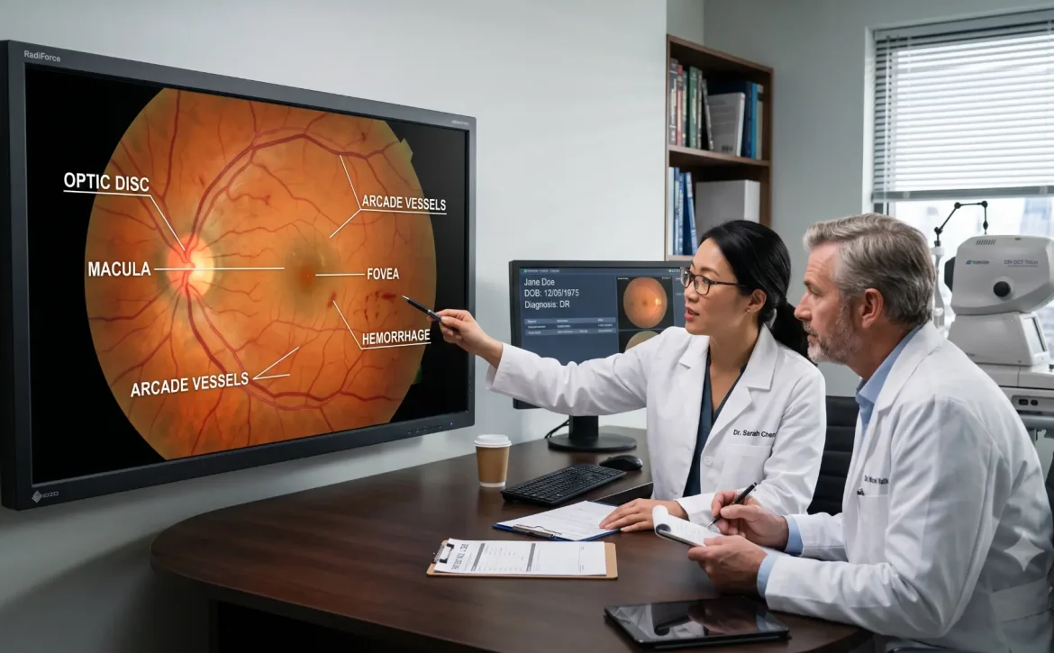
The process begins with high-quality retinal images captured using fundus cameras or OCT systems. Automatic retinal image analysis then uses specialized software to enhance image quality, detect important anatomical structures such as the optic disc, macula, and retinal blood vessels, and identify abnormalities including microaneurysms, hemorrhages, hard exudates, and other retinal lesions. The results of automatic retinal image analysis are presented as clinical decision-support information, enabling eye care professionals to combine the findings with clinical examination and patient history before making a diagnosis or recommending appropriate treatment.
How Does Automatic Retinal Image Analysis Work?
Automatic retinal image analysis follows a structured workflow that converts retinal images into clinically useful information. The process begins with,
- image acquisition, where high-quality retinal images are captured using fundus cameras, optical coherence tomography (OCT), or portable retinal imaging devices. Clear images are essential because image quality directly affects the accuracy of the analysis.
- Next, the software performs image preprocessing by reducing noise, adjusting brightness and contrast, and correcting uneven illumination. These enhancements improve the visibility of retinal structures and help ensure reliable analysis.
- The system then moves to feature detection, identifying important anatomical landmarks such as the optic disc, macula, retinal blood vessels, and retinal nerve fiber layer. It also detects abnormalities such as microaneurysms, hemorrhages, hard exudates, cotton wool spots, and drusen.
- Finally, AI-powered clinical decision-support compares these features with validated clinical patterns and flags images that may require specialist review. Rather than providing a definitive diagnosis, the technology supports clinicians by highlighting suspicious findings and helping prioritize patients for further evaluation.
Conditions Detected Through Automated Retinal Image Analysis
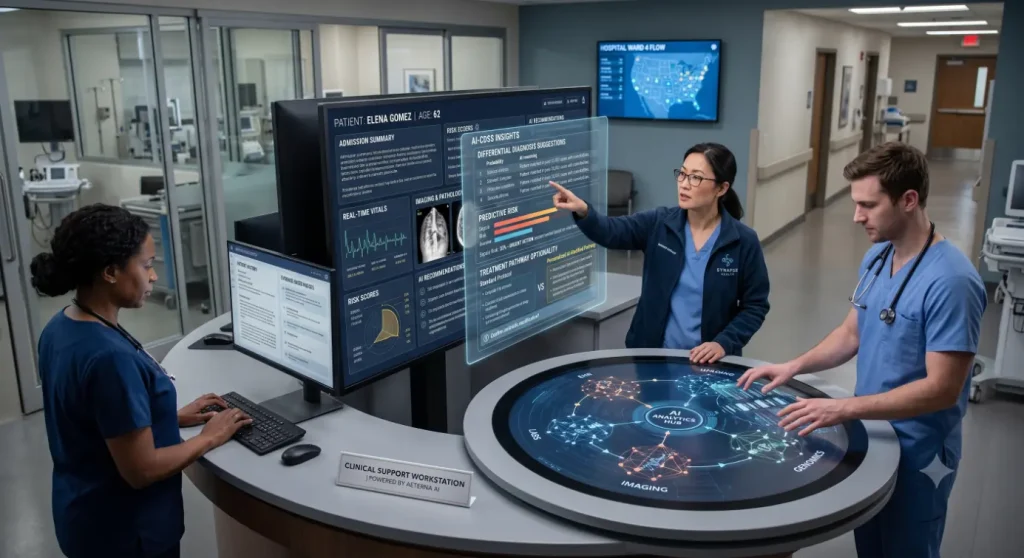
Automated retinal image analysis can support the evaluation of several retinal diseases that benefit from early detection and timely treatment. One of its most common applications is diabetic retinopathy, where it helps identify early signs of retinal blood vessel damage. It can also assist in evaluating age-related macular degeneration (AMD) by detecting drusen and other macular changes associated with disease progression.
In addition, the technology supports screening for glaucoma by assessing optic nerve head characteristics, while diabetic macular edema (DME) may be identified through retinal thickening and fluid-related changes, particularly when combined with OCT imaging. Other conditions that may be evaluated include hypertensive retinopathy, retinal vein occlusion, and certain pathological myopia-related retinal abnormalities. Although these systems improve screening efficiency, all findings should be confirmed through a comprehensive clinical examination by a qualified eye care professional.
Also Read: AI Retinal Image Analysis Tools
Benefits of Automated Retinal Image Analysis
- The adoption of automated retinal image analysis is improving the efficiency and accessibility of retinal disease screening across a wide range of healthcare settings. By reducing the time required to review retinal images, these systems enable eye care professionals to screen more patients while maintaining consistent evaluation standards.
- One of the most significant advantages is earlier detection of retinal diseases. By identifying subtle retinal abnormalities that may otherwise be overlooked during routine screening, automated systems help clinicians prioritize patients who require comprehensive eye examinations. Earlier referrals can support timely treatment, which may reduce the risk of vision loss for many retinal conditions.
- Another important benefit is improved consistency. Unlike manual image interpretation, which can vary between observers, automated retinal image analysis applies standardized evaluation methods to every image. This supports more uniform screening outcomes and enhances quality assurance in clinical practice.
- The technology also strengthens teleophthalmology and remote eye care services. Healthcare providers can analyze retinal images captured in community clinics, primary care centers, or rural healthcare facilities, allowing specialists to review high-priority cases without requiring every patient to travel to a tertiary eye hospital. In addition, AI-powered clinical decision-support helps streamline clinical workflows by reducing the burden of reviewing large image datasets while ensuring that healthcare professionals remain responsible for diagnosis and treatment decisions.
Also Read:Retinal Image Analysis – Easy Detection of Eye Diseases
Automated Analysis of Retinal Images at Population Scale
- The automated analysis of retinal images at population scale is becoming increasingly important as chronic diseases such as diabetes and hypertension continue to rise worldwide, creating a growing need to screen millions of individuals for vision-threatening eye diseases. Automatic retinal image analysis provides a practical solution by enabling large volumes of retinal images to be processed efficiently, consistently, and with the support of AI-powered clinical decision-support. As a result, healthcare systems can improve screening capacity, prioritize high-risk patients for specialist review, and strengthen early detection programs across diverse populations.
- National diabetic eye screening programs, community vision initiatives, mobile eye clinics, and telemedicine networks can benefit from the automated analysis of retinal images at population scale. Powered by automatic retinal image analysis, these systems rapidly identify retinal images that may contain signs of retinal disease, allowing ophthalmologists to focus on patients who require urgent evaluation while reducing the burden of manual image review.
- Beyond clinical care, the automated analysis of retinal images at population scale supports public health planning, epidemiological research, and disease surveillance by providing valuable insights into the prevalence and distribution of retinal disorders. As digital healthcare continues to evolve, automatic retinal image analysis is expected to play an increasingly important role in improving access to preventive eye care, expanding large-scale screening programs, and reducing avoidable vision loss worldwide.
Applications in Modern Healthcare
Automated retinal image analysis is becoming an integral part of modern ophthalmology and preventive eye care. Hospitals and specialty eye clinics use these systems to support routine retinal screening, while primary care providers and diabetes clinics incorporate retinal imaging into regular health assessments for patients at risk of vision-threatening complications. The technology also plays a valuable role in teleophthalmology, enabling retinal images captured in remote or underserved communities to be securely reviewed by eye care specialists. In addition, researchers use automated retinal image analysis to evaluate large imaging datasets, supporting clinical studies and the development of improved screening strategies.
Challenges and Limitations
Despite its many advantages, automated retinal image analysis has certain limitations. Image quality remains one of the most important factors affecting performance, as blurred images, poor lighting, or media opacities can reduce analysis accuracy. Algorithm performance may also vary depending on the imaging device, patient population, and clinical setting. For these reasons, automated systems should always be viewed as AI-powered clinical decision-support rather than standalone diagnostic tools. Qualified eye care professionals must interpret the findings alongside a patient’s medical history, symptoms, and comprehensive eye examination. Data privacy, cybersecurity, regulatory compliance, and continuous clinical validation are also essential for the responsible implementation of these technologies.
The Future of Automated Retinal Image Analysis
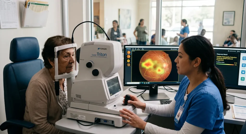
The future of automated retinal image analysis is expected to be driven by advances in artificial intelligence, retinal imaging technology, and digital healthcare infrastructure. Future systems are likely to integrate multiple imaging modalities, including fundus photography and optical coherence tomography (OCT), to provide more comprehensive assessments. Portable retinal cameras, smartphone-based imaging, cloud computing, and secure electronic health record integration may further expand access to retinal screening, particularly in underserved regions. Continued improvements in AI-powered clinical decision-support are expected to enhance screening efficiency while supporting clinicians in delivering timely and evidence-based patient care.
Conclusion
Automated retinal image analysis is transforming retinal screening by improving the speed, consistency, and accessibility of image evaluation. Whether described as automated retinal image analysis or automatic retinal image analysis, these technologies support healthcare professionals in identifying patients who may benefit from further clinical assessment. Furthermore, the automated analysis of retinal images at population scale has the potential to strengthen national screening programs, expand access to preventive eye care, and support better public health outcomes. As research and digital imaging technologies continue to evolve, automated retinal image analysis will remain an important component of modern ophthalmology, working alongside clinical expertise to improve patient care rather than replace it.
Also Read: Retinal Image Analysis Software: Easy Eye Disease Detection
Frequently Asked Questions (FAQs)
1.What is automated retinal image analysis?
Automated retinal image analysis is a computer-assisted process that evaluates retinal images using image-processing techniques and AI-powered clinical decision-support to help healthcare professionals identify potential retinal abnormalities more efficiently.
2.How does automatic retinal image analysis work?
The process involves retinal image acquisition, image preprocessing, feature detection, and AI-assisted analysis. The system highlights suspicious findings that clinicians review before making diagnostic and treatment decisions.
3.Which eye diseases can automated retinal image analysis help evaluate?
It can support the evaluation of diabetic retinopathy, age-related macular degeneration (AMD), glaucoma, diabetic macular edema, hypertensive retinopathy, retinal vein occlusion, and other retinal abnormalities.
4.What is automated analysis of retinal images at population scale?
It refers to the use of automated systems to evaluate large numbers of retinal images collected through national screening programs, teleophthalmology services, and community eye care initiatives, helping healthcare providers identify patients who may require specialist evaluation more efficiently.
5.Can automated retinal image analysis replace ophthalmologists?
No. Automated retinal image analysis is designed to assist healthcare professionals by improving screening efficiency and supporting clinical decision-making. Final diagnosis, treatment planning, and patient management remain the responsibility of qualified ophthalmologists and other eye care professionals.
Also Read:Conditions Predicted by Retinal Image Analysis