Retinal image analysis applications plays a vital role in modern eye care, helping ophthalmologists assess retinal health accurately and efficiently. The retina contains a complex network of blood vessels, nerves, and light-sensitive tissues that often show the earliest signs of eye diseases as well as certain systemic health conditions. By examining retinal images captured through technologies such as fundus photography and optical coherence tomography (OCT), clinicians can detect subtle abnormalities before noticeable vision loss occurs. The growing use of artificial intelligence (AI), deep learning, and advanced image processing has further improved retinal image analysis.
Key Takeaways
- Detects retinal diseases earlier for timely treatment.
- AI improves the speed and accuracy of retinal screening.
- Retinal imaging may help identify signs of systemic diseases.
- The AI-powered retina image analysis market is growing rapidly.
Intelligent software can automatically identify suspicious features, measure retinal structures, and assist ophthalmologists during diagnosis. As the number of people affected by diabetes, glaucoma, and age-related eye diseases continues to rise, retinal image analysis has become an important tool for early detection, routine screening, disease monitoring, and treatment planning. Rather than replacing medical professionals, these technologies provide valuable clinical support that helps improve decision-making and patient care.
What Is Retinal Image Analysis Applications?
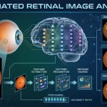
Retinal image analysis applications is the process of examining digital retinal images to evaluate eye health and identify abnormalities. Images are typically captured using fundus cameras or optical coherence tomography (OCT) devices, allowing specialists to examine key retinal structures such as the optic disc, macula, retinal blood vessels, and nerve fiber layers. Modern retinal image analysis applications use image enhancement, automated measurements, and AI-based pattern recognition to assist healthcare professionals in detecting abnormalities, assessing disease severity, and generating decision-support reports.
These technologies reduce manual workload while improving the accuracy and consistency of retinal examinations. Researchers are also exploring AI retinal imaging for full body health analysis to investigate whether retinal biomarkers can help identify certain systemic health conditions, although this field is still evolving. As healthcare providers increasingly adopt AI-driven diagnostic solutions, the AI-powered retina image analysis market continues to grow, driven by the demand for faster screening, improved clinical efficiency, and more accessible eye care services.
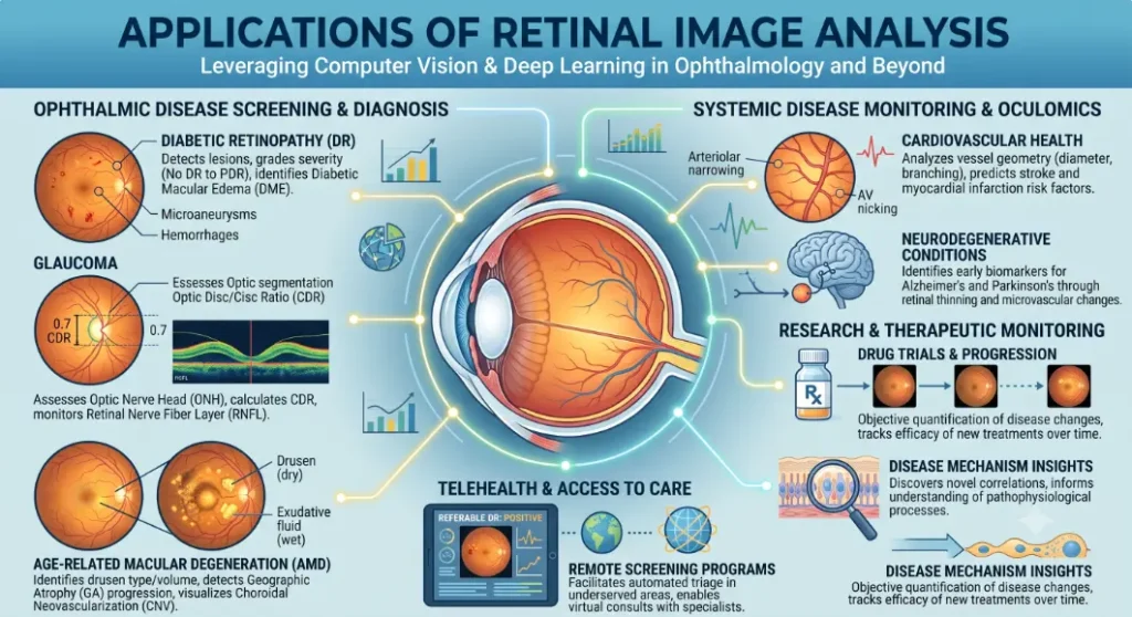
Also Read: Retinal Image Analysis – Easy Detection of Eye Diseases
Major Applications of Retinal Image Analysis Applications
1.Diabetic Retinopathy Screening
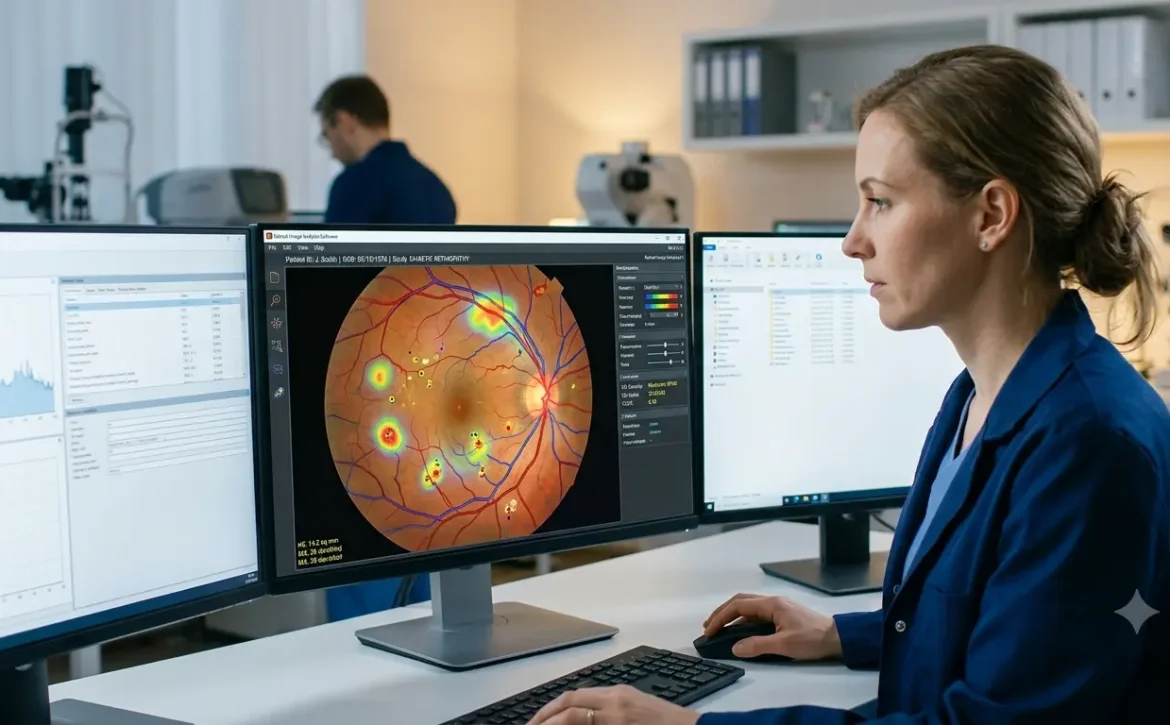
Diabetic retinopathy is one of the most common causes of preventable blindness among people with diabetes. Since the disease often develops without noticeable symptoms, routine retinal examinations are critical. Retinal image analysis applications helps detect early signs such as microaneurysms, retinal hemorrhages, hard exudates, and cotton wool spots. Automated screening systems enable healthcare providers to evaluate large numbers of retinal images quickly, making diabetic eye screening more accessible and efficient while ensuring patients with advanced disease receive timely specialist care.
2.Glaucoma
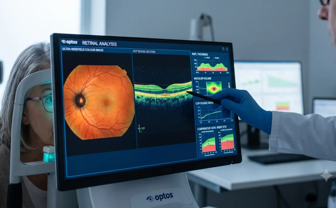
Assessment Glaucoma gradually damages the optic nerve and frequently progresses without obvious symptoms until irreversible vision loss has occurred. Early diagnosis is therefore essential. Retinal image analysis applications evaluates the optic nerve head, measures the cup-to-disc ratio, and analyzes retinal nerve fiber layer thickness using OCT images. Comparing these measurements over multiple visits helps clinicians identify disease progression and assess how well treatment is controlling the condition.
3.Age-related macular degeneration (AMD)
Age-related macular degeneration (AMD) affects the macula, the part of the retina responsible for sharp, central vision. Detecting AMD at an early stage can improve long-term visual outcomes. Image analysis assists ophthalmologists in identifying drusen, pigment abnormalities, retinal fluid, and areas of geographic atrophy. Regular imaging helps ophthalmologists monitor disease progression over time and evaluate how well a treatment is working.
4.Retinal Vascular Disease
EvaluationChanges in the retina’s blood vessels can indicate conditions such as retinal vein occlusion, retinal artery occlusion, and hypertensive retinopathy.Retinal image analysis applications enhances blood vessel visibility, measures vascular features, and detects subtle abnormalities that may be missed during routine examinations. This supports earlier diagnosis and more effective treatment planning. Advanced image analysis enhances vessel visibility, measures vascular characteristics, and identifies subtle abnormalities that may not be easily recognized during routine examination. These measurements support earlier diagnosis and better management of retinal vascular diseases.
5.Macular Edema Monitoring
Macular edema occurs when excess fluid collects in the macula, reducing the sharpness of central vision. It is commonly associated with diabetic retinopathy and retinal vascular diseases. Today’s retinal image analysis applications use OCT imaging and AI algorithms to detect fluid accumulation, measure retinal thickness, and provide precise, data-driven insights for treatment planning. Beyond eye disease management, researchers are investigating AI retinal imaging for full body health analysis to explore whether retinal changes can help identify early signs of cardiovascular, metabolic, and neurological conditions. These ongoing innovations are also fueling the rapid growth of the AI-powered retina image analysis market, as healthcare providers increasingly adopt intelligent imaging solutions for faster diagnosis and improved patient outcomes.
6.Monitoring Inherited Retinal Disorders
Inherited retinal diseases such as retinitis pigmentosa and Stargardt disease often require lifelong observation. Retinal image analysis provides a consistent method for documenting structural retinal changes and comparing disease progression over time. It also plays an important role in clinical research by helping investigators assess the effectiveness of emerging therapies.
7.Applications Beyond Eye Diseases
Researchers are increasingly exploring retinal imaging as a non-invasive method for assessing overall health. Because retinal blood vessels can be directly observed, they may provide valuable information about systemic conditions including diabetes, hypertension, and cardiovascular disease. Scientists are also investigating retinal biomarkers associated with neurological disorders such as Alzheimer’s disease and Parkinson’s disease. Although these applications remain under active research, they demonstrate the expanding clinical value of retinal imaging beyond traditional ophthalmology.
Benefits of Retinal Image Analysis Applications
Retinal image analysis applications offers numerous advantages for healthcare professionals and patients by combining advanced imaging technologies with AI-assisted clinical support.
- Detects retinal abnormalities at earlier stages.
- Improves the accuracy and consistency of image interpretation.
- Supports timely diagnosis of vision-threatening diseases.
- Reduces the workload associated with reviewing large image datasets.
- Enables long-term monitoring of disease progression.
- Strengthens diabetic retinopathy screening programs.
- Improves workflow efficiency in busy ophthalmology clinics.
- Supports telemedicine and remote eye screening services.
Also Read: Retinal Image Analysis Software: Easy Eye Disease Detection
Challenges and Limitations
Despite its many benefits, retinal image analysis also has certain limitations. The accuracy of the results depends heavily on image quality. Factors such as cataracts, poor focus, inadequate illumination, or patient movement can reduce image clarity and affect the analysis. While AI retinal imaging for full body health analysis is an emerging area of research, these applications are still under investigation and should not be considered a replacement for established diagnostic methods. AI-generated findings should always be interpreted alongside clinical examination, patient history, and professional medical judgment. Healthcare organizations must also address data privacy, regulatory compliance, and system integration to ensure safe and effective implementation.
AI Retinal Imaging for Full Body Health Analysis
Researchers are exploring AI retinal imaging for full body health analysis to determine whether retinal images can reveal early signs of systemic diseases such as diabetes, hypertension, cardiovascular disease, and certain neurological disorders. These emerging retinal image analysis applications have the potential to support earlier health risk assessment through a simple, non-invasive eye examination. Although this technology is still undergoing clinical validation, its growing potential is contributing to the rapid expansion of the AI-powered retina image analysis market, where AI is increasingly being used to enhance both ophthalmic and systemic disease screening.
AI-Powered Retina Image Analysis Market
The AI-powered retina image analysis market is experiencing rapid growth as healthcare providers increasingly adopt AI-driven solutions for early disease detection and large-scale retinal screening. Modern retinal image analysis applications are helping hospitals, diagnostic centers, and teleophthalmology programs improve diagnostic accuracy, reduce screening time, and expand access to quality eye care. As research into AI retinal imaging for full body health analysis continues to advance, the market is expected to grow further, creating new opportunities for both ophthalmic and broader healthcare diagnostics.

Future Trends
Advances in artificial intelligence, deep learning, and retinal imaging technologies are expected to expand the capabilities of retinal image analysis applications in the coming years. Future systems may detect eye diseases earlier, predict disease progression more accurately, and combine data from multiple imaging techniques to support better clinical decisions. Researchers are also exploring AI retinal imaging for full body health analysis to identify potential biomarkers associated with systemic conditions such as cardiovascular and neurological diseases, although these applications are still under investigation.
In addition, the growing adoption of AI solutions, cloud-based platforms, and teleophthalmology is contributing to the rapid growth of the AI-powered retina image analysis market, improving access to specialist eye care, particularly in underserved and rural communities. As these technologies continue to evolve, retinal image analysis application will play an increasingly important role in preventive healthcare and personalized treatment planning.
Conclusion
Retinal image analysis applications has become an indispensable technology in modern ophthalmology. It supports clinicians by improving the detection, diagnosis, and monitoring of a wide range of retinal diseases, including diabetic retinopathy, glaucoma, macular degeneration, retinal vascular disorders, and inherited retinal conditions. As AI and imaging technologies continue to advance, retinal image analysis will play an increasingly important role in delivering faster, more accurate, and more accessible eye care. While intelligent software enhances clinical efficiency, the expertise of qualified ophthalmologists remains essential for accurate diagnosis, treatment decisions, and comprehensive patient management.
Frequently Asked Questions
1. What is retinal image analysis applications?
Retinal image analysis is the computerized evaluation of retinal images to identify abnormalities, measure retinal structures, and assist eye care professionals in diagnosing and monitoring eye diseases.
2. Which eye conditions can retinal image analysis applications detect?
It is widely used to support the detection of diabetic retinopathy, glaucoma, age-related macular degeneration, macular edema, retinal vascular diseases, and inherited retinal disorders.
3. How does AI contribute to retinal image analysis applications ?
AI algorithms can automatically identify disease-related features, perform retinal measurements, highlight suspicious areas, and provide decision-support information for clinicians.
4. Can retinal image analysis replace ophthalmologists?
No. These technologies are designed to support clinical decision-making. Diagnosis and treatment should always be confirmed by qualified eye care professionals.
5. Is retinal image analysis useful beyond ophthalmology?
Yes. Researchers are studying whether retinal imaging can help identify biomarkers associated with diabetes, cardiovascular disease, hypertension, and certain neurological disorders, although many of these applications are still under investigation.
6. Which imaging technologies are commonly used for retinal image analysis applications?
The most commonly used imaging technologies include fundus photography, optical coherence tomography (OCT), OCT angiography (OCTA), and ultra-widefield retinal imaging, all of which provide detailed views of the retina for accurate examination and diagnosis.
1. What is AI retinal imaging for full body health analysis?
It uses AI to analyze retinal images for signs of systemic diseases such as diabetes, hypertension, and cardiovascular conditions, supporting early risk assessment.
2. Why is the AI-powered retina image analysis market growing?
The market is growing due to increasing demand for early disease detection, AI-assisted retinal screening, and wider adoption of teleophthalmology and automated retinal image analysis applications.
Also Read: Conditions Predicted by Retinal Image Analysis