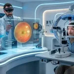
Early detection of retinal diseases with AI assisted diagnosis is the application of data-driven diagnostic systems to identify retinal abnormalities before clinical symptoms become severe. This approach enhances screening efficiency and diagnostic precision in ophthalmology. Can early intervention significantly reduce blindness rates? Research data confirms that timely identification is essential in maintaining visual health.
Key Takeaways
- Early detection significantly reduces the risk of vision loss
- AI-assisted systems improve diagnostic speed and consistency
- Standard imaging methods enable scalable screening
- Regulatory compliance is essential for clinical deployment
- Integration challenges remain but are actively being addressed
What is early detection of retinal diseases with AI assisted diagnosis?
Early detection of retinal diseases with AI assisted diagnosis involves automated analysis of retinal images to identify pathological changes.
Key characteristics:
- Uses fundus photography and OCT (Optical Coherence Tomography)
- Detects microvascular and structural abnormalities
- Supports clinicians with probability-based outputs
Common diseases detected:
- Diabetic retinopathy (DR)
- Age-related macular degeneration (AMD)
- Glaucoma-related optic nerve damage
How does early detection of retinal diseases with AI assisted diagnosis work?
Early detection of retinal diseases with AI assisted diagnosis works by processing medical images through trained systems that recognize disease patterns.
Typical workflow:
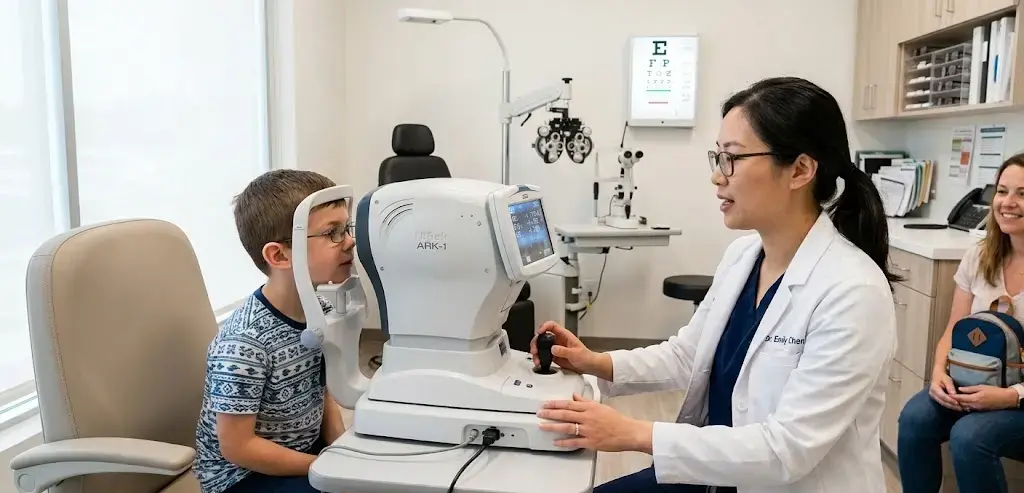
- Image acquisition (fundus camera or OCT)
- Image preprocessing (noise reduction, normalization)
- Feature extraction (lesions, vessels, macula)
- Classification and risk scoring
- Clinical interpretation
Example:
Automated retinal disease assessment (ARDA) systems classify diabetic retinopathy severity based on standardized grading scales such as ETDRS.
What are the clinical benefits of early detection?
Early detection of retinal diseases with AI assisted diagnosis provides measurable improvements in patient care.
Key benefits:
- Earlier intervention reduces vision loss risk
- Increased screening coverage in underserved areas
- Consistent diagnostic accuracy across populations
- Reduced clinician workload
Clinical comparison:
| Parameter | Traditional Screening | AI-Assisted Detection |
| Detection Speed | Moderate | High |
| Consistency | Variable | Standardized |
| Accessibility | Limited | Scalable |
| Error Rate | Higher | Lower |
Which technologies are used in retinal disease detection?
Early detection of retinal diseases with AI assisted diagnosis relies on multiple imaging and computational technologies.
Core technologies:
- Fundus imaging systems
- Optical Coherence Tomography (OCT)
- Deep image classification models
- Cloud-based diagnostic platforms
A related area often discussed includes automated retinal screening systems, retinal image analysis tools, and AI-based ophthalmology platforms.
What are the regulatory and clinical standards?
Early detection of retinal diseases with AI assisted diagnosis must comply with strict medical and regulatory frameworks.
Key standards:
- FDA approval for diagnostic software (USA)
- CE marking under MDR (Europe)
- ISO 13485 for medical device quality
- Clinical validation through peer-reviewed trials
Classification:
- Screening tools (risk identification)
- Diagnostic support systems
- Autonomous diagnostic systems (limited approval cases)
What are the challenges and limitations?
Early detection of retinal diseases with AI assisted diagnosis faces several implementation challenges.
Primary limitations:
- Data bias affecting diagnostic fairness
- Dependence on image quality
- Integration with hospital systems
- Regulatory approval complexity
Operational concern:
Variability in imaging conditions can reduce detection reliability in real-world settings.
Conclusion
Early detection of retinal diseases with AI assisted diagnosis is reshaping ophthalmic screening by improving accuracy, accessibility, and clinical efficiency. Its structured classification into screening, support, and autonomous systems ensures clear implementation pathways. As healthcare systems evolve, this approach aligns closely with broader advancements such as faster and more accurate patient visits with AI assisted diagnosis, enabling more efficient and proactive care delivery.
FAQs
What diseases can be detected early using AI-assisted retinal diagnosis?
Screening methods typically detect diabetic retinopathy, macular degeneration, and glaucoma at initial stages.
Is AI-assisted retinal diagnosis clinically reliable?
Yes, many systems demonstrate high sensitivity and specificity when validated in clinical studies.
What imaging methods are used in retinal diagnosis?
Fundus photography and Optical Coherence Tomography (OCT) are the primary imaging techniques.
Are AI-based retinal tools approved by regulators?
Some systems have received FDA approval or CE marking after clinical validation.
Can AI replace ophthalmologists in retinal diagnosis?
No, these systems support clinical decisions but do not replace professional judgment.
Sources
https://pmc.ncbi.nlm.nih.gov/articles/PMC11052176/
https://www.mdpi.com/2075-4426/14/7/690
https://pubmed.ncbi.nlm.nih.gov/38674173/
https://www.retinaconsultantstexas.com/blog/the-role-of-artificial-intelligence-in-retinal-disease-diagnosis
https://www.sciencedirect.com/science/article/pii/S2949953424000109
https://onlinelibrary.wiley.com/doi/10.1155/ijbi/6154285
https://macretinahospital.com/the-role-of-ai-in-diagnosing-retinal-disorders-at-an-early-stage/
https://www.nature.com/articles/s41591-026-04359-w
https://www.laxmieye.org/blog/ai-eye-care-early-detection-revolution/
https://www.frontiersin.org/journals/cell-and-developmental-biology/articles/10.3389/fcell.2022.1053483/full