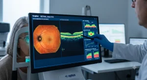
Retinal imaging is increasingly used in large-scale eye screening. However, reviewing thousands or millions of images can be difficult. Automated Analysis of Retinal Images at Population Scale helps process large volumes of retinal images, identify relevant features, and prioritize cases for clinical review.
Key Takeaways
- Automated analysis helps process large volumes of retinal images efficiently.
- It can support screening for conditions such as diabetic retinopathy and glaucoma.
- Image quality assessment can identify images that need to be recaptured.
- Clinical review remains important for abnormal or uncertain findings.
This is where automated analysis of retinal images at population scale can be useful. Computer-based image analysis can examine retinal photographs, identify specific features, check image quality, and help determine which cases may need closer clinical attention.
Why Automated Analysis of Retinal Images at Population Scale Analysis Matters?
In a small eye clinic, a specialist may be able to review each retinal image manually. That becomes much harder when screening is conducted across several hospitals, community centers, diabetes clinics, or rural healthcare facilities.
Large screening programs can face several practical problems:
- A high number of retinal images to review
- Limited availability of trained eye-care professionals
- Poor-quality or ungradable images
- Differences in manual assessment
- Delays in referring patients for further examination
- Difficulty managing and organizing large imaging datasets
Automated analysis can perform an initial assessment of images before they reach the next stage of the clinical workflow.
Also Read: Software for Automating Medical Practice Workflows
How Automated Retinal Image Analysis Works
The exact process depends on Automated Analysis of Retinal Images at Population Scale may include several steps.
| Stage | What happens |
|---|---|
| Image capture | A retinal camera captures fundus photographs |
| Quality check | The system assesses whether the image is suitable for analysis |
| Image processing | Relevant retinal structures are identified and processed |
| Feature detection | Potential abnormalities or measurable features are identified |
| Classification | Images may be placed into predefined screening categories |
| Clinical review | Cases requiring further assessment are reviewed by healthcare professionals |
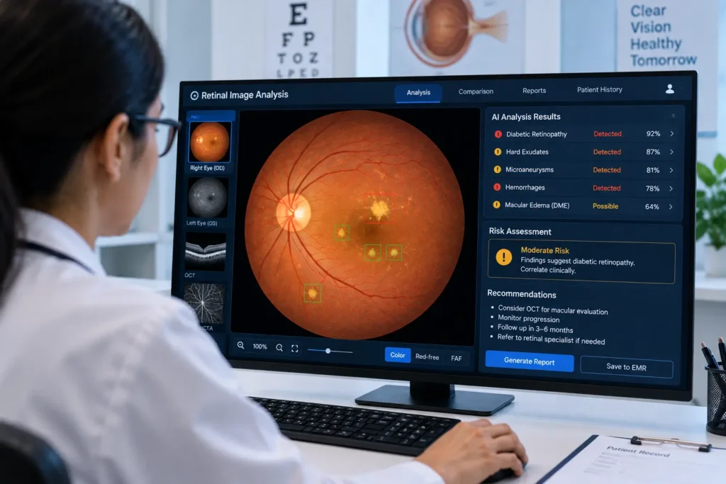
For example, an automated system may identify retinal features associated with diabetic retinopathy, such as microaneurysms or hemorrhages. Other systems may examine the optic disc when supporting glaucoma screening.
The output depends on how the system has been developed and validated. Automated findings should be interpreted within the appropriate clinical and regulatory framework.
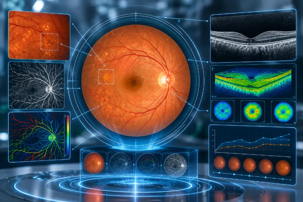
Conditions That Can Be Assessed
Retinal image analysis has been studied across several areas of eye care.
Diabetic retinopathy: Retinal photographs can be analyzed for lesions such as microaneurysms, hemorrhages, and exudates.
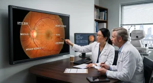
Glaucoma: Analysis may include measurements or characteristics of the optic disc and optic cup that are relevant to glaucoma assessment.
Age-related macular degeneration: Systems may examine the macular region for features such as drusen and other retinal changes.
Retinal vascular changes: Automated vessel analysis can measure characteristics such as vessel width, branching, and tortuosity.
The performance of an automated system can vary depending on image quality, camera type, patient population, and the specific task for which it was validated.
Where Population-Scale Analysis Can Be Used
Automated Analysis of Retinal Images at Population Scale can be useful in different healthcare settings, including:
- National or regional eye-screening programs
- Diabetes screening initiatives
- Primary healthcare centers
- Community eye-care programs
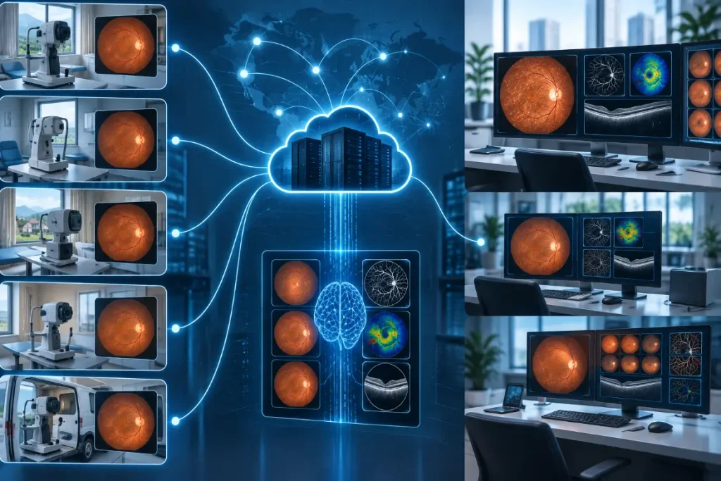
- Mobile eye clinics
- Teleophthalmology services
- Research and population-health studies
In a community screening program, for example, retinal photographs could be captured locally and processed through an analysis platform. Images that require additional attention could then be referred to an appropriate eye-care professional.
Benefits and Challenges
The potential benefits are mainly related to handling large amounts of information consistently.
Potential benefits include:
- Faster initial assessment of large image collections
- Support for screening and patient prioritization
- More consistent application of predefined analysis criteria
- Identification of ungradable images
- Better organization of large retinal datasets
- Support for remote screening programs
However, Automated Analysis of Retinal Images at Population Scale also has limitations. Different cameras can produce different image characteristics, and performance can change across patient populations. Poor image quality can also affect results.
Privacy and cybersecurity are important when large numbers of patient images are collected and transferred. Healthcare organizations also need to consider regulatory requirements, clinical validation, data governance, and appropriate human oversight.
The Future of Automated Retinal Image Analysis
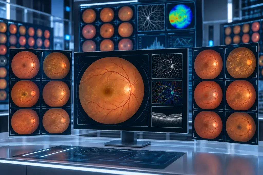
Population-scale retinal screening is likely to involve several technologies rather than a single imaging method. Fundus photography may be combined with OCT, OCTA, or ultra-widefield imaging when clinically appropriate.
As computational retinal image analysis develops, systems may provide more detailed measurements and support longitudinal monitoring of retinal changes. However, wider adoption will depend on evidence showing that these tools work reliably in the populations and healthcare settings where they are used.
The most practical role for automation is to support clinicians with large volumes of retinal information while keeping clinical judgment at the center of patient care.
Also Read: Future-Proof Medical Software
Frequently Asked Questions
What is automated analysis of retinal images?
It is the use of computer-based image analysis to examine retinal photographs, identify specific features, assess image quality, and support screening or clinical decision-making.
Why is automated analysis useful for population screening?
Large screening programs can generate more retinal images than specialists can easily review manually. Automation can help process these images and identify cases that may need additional attention.
Can automated retinal analysis diagnose eye diseases?
Some systems are designed and validated for specific screening or diagnostic-support tasks. Their results should be interpreted according to their intended use and alongside appropriate clinical assessment.
Does automated retinal analysis replace ophthalmologists?
No. It is generally used as a support tool. Patients with abnormal, uncertain, or ungradable results may require examination and assessment by qualified eye-care professionals.
What types of retinal imaging can be analyzed?
Fundus photography is widely used for retinal image analysis. Depending on the application, other modalities such as OCT, OCTA, and ultra-widefield retinal imaging may also be analyzed.