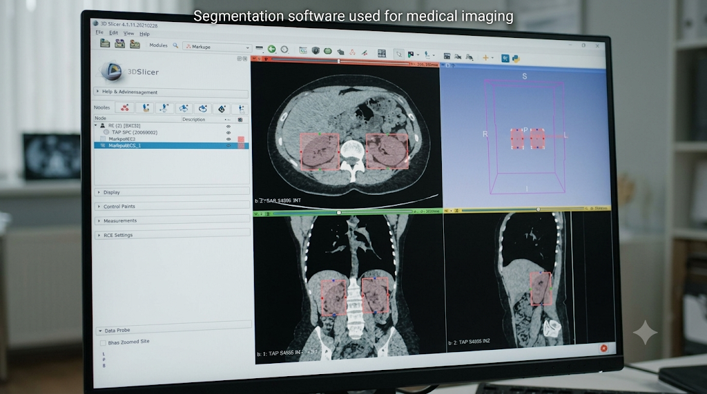
Segmentation software used for medical imaging turns raw CT, MRI, and ultrasound scans into labeled structures such as organs, bones, vessels, and tumors. Rather than viewing an indistinct blob of pixels, clinicians and engineers receive sharp boundaries that they can quantitatively analyze, visually inspect, and use to plan procedures. With increases in scan volume and advances in automation, segmentation has become an indispensable component of diagnostic imaging, surgical planning, radiotherapy treatment planning, and biomedical device design. This guide outlines the process, tools, and considerations required to perform segmentation safely.
Key Takeaways
- Each structure in a scan receives a label in segmentation, allowing for quantification and modeling.
- With deep learning, segmentation has gotten faster, although results require expert analysis.
- Validation, bias evaluation, and regulatory strategy are just as important as segmentation accuracy.
Table of Contents
Segmentation Software Used for Medical Imaging: How It Works
A medical scan is a grid of pixels (2D) or voxels (3D), each holding an intensity value. Segmentation assigns each one to a category, such as bone, soft tissue, or lesion. The output is a mask or label map that can be turned into measurements or a 3D model.
A typical workflow has four stages:
- Import: Scans are loaded in standard formats such as DICOM or NIfTI.
- Preprocessing: Noise is reduced, images are aligned, and intensities are normalized so scans from different machines look comparable.
- Segmentation: The software outlines the target structures.
- Review and analysis: A clinician corrects errors, then calculates volume, shape, or other measurements.
Skipping the review step is where many real-world problems begin, which is why human oversight remains essential.
Main Segmentation Methods
| Method | How It Works | Best For |
| Thresholding | Separates regions by intensity value | High-contrast structures like bone on CT |
| Region growing | Starts from a seed point and expands to similar neighbors | Connected, uniform structures |
| Clustering | Groups pixels with similar features, for example K-means | Tissue types with distinct texture or color |
| Edge and contour methods | Follows boundaries where intensity changes sharply | Well-defined organ outlines |
| Atlas-based | Maps a labeled reference anatomy onto a new scan | Brain and standard anatomy |
| Deep learning | Neural networks learn to label every pixel from examples | Complex anatomy and large datasets |
Classical methods are transparent and fast but can struggle with noisy or low-contrast images. Deep learning handles variation better, though it needs high-quality training data and careful testing.
Common Tools and Platforms
Teams choose from open-source projects, commercial packages, and programming environments:
- 3D Slicer and ITK-SNAP: Widely used open-source applications for viewing and segmenting images, popular in research.
- MONAI: An open-source framework for building and training medical imaging AI models.
- Simpleware (Synopsys/Ansys) and Materialise Mimics: Commercial software for turning scans into 3D models for surgical planning, implants, and simulation.
- MATLAB Medical Imaging Toolbox: A programming environment with apps for labeling, preprocessing, and building segmentation algorithms.
- Vendor-integrated tools: Scanner and PACS makers often include their own segmentation features.
Many commercial platforms now offer automated modules for specific anatomy, such as hips, knees, or the heart, while leaving manual cleanup available when images are noisy or pathology is unusual.
Segmentation Software Used for Medical Imaging – Key Application Areas
- Surgical planning: Patient-specific 3D models help surgeons rehearse complex procedures.
- Radiotherapy: Accurate outlines of tumors and nearby organs guide radiation dose.
- Device and implant design: Engineers use real anatomy to design guides and implants.
- Disease monitoring: Tracking tumor volume or lung changes over time.
- Research and virtual trials: Large cohorts can be analyzed or simulated at scale.
- 3D printing: Anatomical models support teaching and preoperative preparation.
Vendors report that automated segmentation can cut the time needed for some tasks from hours to minutes, which frees specialists for higher-value work.
Accuracy Is Not the Whole Story
Teams often judge segmentation with overlap scores such as the Dice coefficient, which compares the software’s outline with an expert’s. A high score is useful but incomplete. Consider:
- Generalization: Does it perform well on scanners, protocols, and patient groups it was not trained on?
- Edge cases: How does it behave with implants, artifacts, or unusual anatomy?
- Bias: Are results consistent across age, sex, body type, and ethnicity?
- Traceability: Can you reproduce a result and see which software version produced it?
- Human factors: Do users over-trust outputs that look polished but are wrong?
Risk Management and Regulation
When segmentation outputs influence diagnosis, treatment, or device manufacturing, the software may be regulated as a medical device. Developers and hospitals should plan for:
- Risk analysis: Identify hazards such as missed tumor boundaries and define controls, following ISO 14971 principles.
- Software lifecycle quality: Document requirements, testing, and changes in line with IEC 62304.
- Cybersecurity and privacy: Protect patient images and follow HIPAA requirements.
- Change control: Retraining or updating an AI model can change its behavior, so updates need verification.
- Post-market monitoring: Track real-world performance and capture incident reports.
Building these steps in early while implementing segmentation software used for medical imaging is far less costly than retrofitting them after launch.

How to Choose Segmentation Software
Ask these questions before committing:
- Which modalities and anatomy do we need, and is the tool validated for them?
- Can clinicians easily review and correct results?
- Does it integrate with our PACS, DICOM workflow, and research tools?
- What evidence supports its accuracy, and on what kinds of data?
- How are updates, audit trails, and access controls handled?
- What regulatory support does the vendor provide?
A small pilot using your own scans will reveal more than a brochure.
The Bottom Line
Segmentation software has become a foundation of modern imaging, turning scans into measurable, actionable information. Automation and deep learning are making it faster and more accessible, but speed alone does not guarantee safety. The strongest programs pair capable tools with clear validation, human review, and structured risk management. Choose software that fits your clinical purpose, test it on your own data, and keep monitoring performance after deployment.
Building or deploying imaging software? Contact Rceenetworks to manage risk and compliance with confidence.
Frequently Asked Questions
1. What is segmentation in medical imaging?
It is the process of dividing a scan into regions, such as organs, tissue types, or lesions, so each can be identified, measured, and modeled.
2. Which software is used for medical image segmentation?
Common options include 3D Slicer, ITK-SNAP, MONAI, Simpleware, Materialise Mimics, and MATLAB tools. The best choice depends on your use case, budget, and regulatory needs.
3. Is AI better than manual segmentation?
AI is usually much faster and can be more consistent, but it can fail on unusual cases. Best practice is automated segmentation followed by expert review.
4. Is medical image segmentation software regulated by the FDA?
It can be, particularly when it is used for diagnosis or treatment decisions. The FDA’s list of AI-enabled medical devices shows how such products are reviewed and authorized.
Also Read: What is Medical Software?